Article Snippet
"Sections were incubated with rabbit anti-mouse laminin polyclonal primary antibody (1:1,000, Sigma-Aldrich, St Louis, MO, USA, #L9393) at 4°C overnight followed by incubation with Alexa Fluor 647-conjugated goat anti-rabbit IgG secondary antibody (1:200, Thermo Fisher Scientific, Waltham, MA, USA, #A32733) at room temperature for 30 min. For myofibroblast identification, sections were incubated with AlexaFluor 647 conjugated anti-mouse αSMA monoclonal antibody (1:100, clone 1A4, Santa Cruz Biotechnology, Dallas, TX, USA, #sc-32251) at 4°C overnight."
Figure Legend
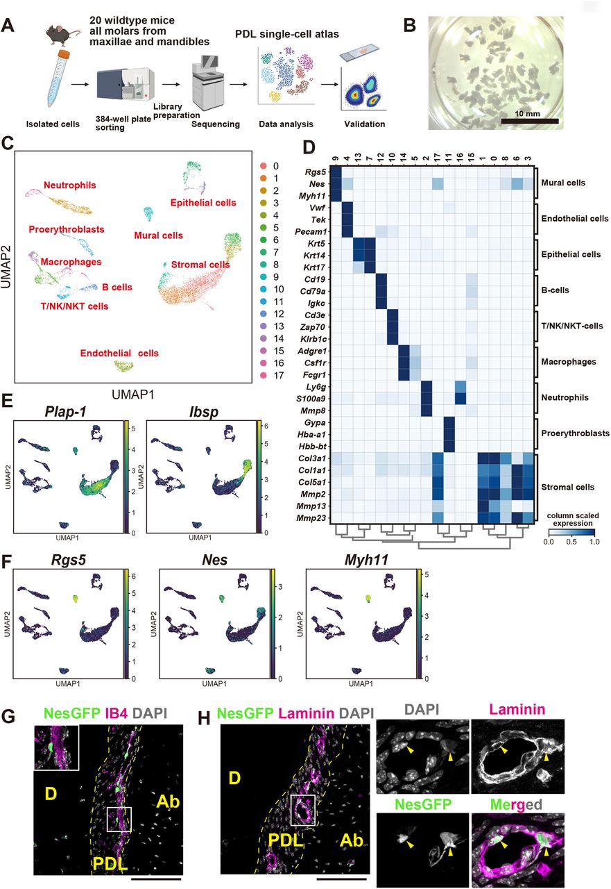
"Figure 4. Single-cell transcriptomic analysis of PDL-derived cells A. Overview of the experimental workflow. Teeth from 20 wild-type mice were subjected to cell isolation, and each cell type was sorted into 384 well plates with a lysis solution. Each lysate was then processed for library preparation, followed by sequencing and bioinformatic analysis. The identified molecules were validated using flow cytometry or histological analysis. B. Representative photograph of extracted teeth used for scRNA-seq analysis. All molars were extracted from dissected maxillae and mandibles. C. scRNA-seq data obtained from 7,318 PDL-derived cells. Clustering analysis with 17 cell types is shown in UMAP. D. A heatmap of the expression of representative genes for each cluster. Each cell type was annotated using these marker genes. E. UMAP plots showing Plap-1 and Ibsp expression. The expression of Plap-1 and Ibsp was mutually exclusive. F. UMAP plots showing Rgs5, Nes , and Myh11 expression. The expressions of Rgs5, Nes , and Myh11 were restricted to mural cell cluster. G. Representative GFP expression and GS-IB-4 staining in the PDL of Nestin-GFP mice. Insets showed Nestin-GFP + cells located in the perivascular area in the PDL. H.Representative laminin immunofluorescent staining in the PDL of Nestin-GFP mice. Nestin-GFP + cell bodies were encapsulated by the endothelial basement membrane. D: Dentin, Ab: Alveolar bone, scale bars: 100 μm. "