Review


Structured Review

Proteintech atg12
Effects of iTBS on the expression levels of autophagy and lysosome-related proteins. A – G Representative protein blots of LC3II/I, SQSTM1, ATG5, <t>ATG12,</t> CTSD, and LAMP1. Normalized to GAPDH. n = 3/group, ns indicates no statistical significance, * P < 0.05, ** P < 0.01, *** P < 0.001, # P < 0.05, ## P < 0.01, ### P < 0.001. Data are expressed as mean ± standard deviation
Atg12, supplied by Proteintech, used in various techniques. Bioz Stars score: 93/100, based on 57 PubMed citations. ZERO BIAS - scores, article reviews, protocol conditions and more
https://www.bioz.com/result/atg12/product/Proteintech
Average 93 stars, based on 57 article reviews
atg12 - by Bioz Stars, 2026-03
93/100 stars

Images

1) Product Images from "Mechanisms of Intermittent Theta-Burst Stimulation Upregulates TFEB and Restores Autophagy to Play a Neuroprotective Role in the Acute Phase After Cerebral Ischemia Reperfusion"

Article Title: Mechanisms of Intermittent Theta-Burst Stimulation Upregulates TFEB and Restores Autophagy to Play a Neuroprotective Role in the Acute Phase After Cerebral Ischemia Reperfusion

Journal: Neurochemical Research

doi: 10.1007/s11064-025-04615-4

Effects of iTBS on the expression levels of autophagy and lysosome-related proteins. A – G Representative protein blots of LC3II/I, SQSTM1, ATG5, ATG12, CTSD, and LAMP1. Normalized to GAPDH. n = 3/group, ns indicates no statistical significance, * P < 0.05, ** P < 0.01, *** P < 0.001, # P < 0.05, ## P < 0.01, ### P < 0.001. Data are expressed as mean ± standard deviation
Figure Legend Snippet: Effects of iTBS on the expression levels of autophagy and lysosome-related proteins. A – G Representative protein blots of LC3II/I, SQSTM1, ATG5, ATG12, CTSD, and LAMP1. Normalized to GAPDH. n = 3/group, ns indicates no statistical significance, * P < 0.05, ** P < 0.01, *** P < 0.001, # P < 0.05, ## P < 0.01, ### P < 0.001. Data are expressed as mean ± standard deviation

Techniques Used: Expressing, Standard Deviation

Effects of iTBS and rTMS on autophagy and lysosome-related protein levels. A – G Representative protein blots of LC3II/I, SQSTM1, ATG5, ATG12, CTSD, LAMP1. Normalized to GAPDH. n = 4/group, ns indicates no statistical significance, * P < 0.05, ** P < 0.01, *** P < 0.001, ## P < 0.01, ### P < 0.001. Data are expressed as mean ± standard deviation
Figure Legend Snippet: Effects of iTBS and rTMS on autophagy and lysosome-related protein levels. A – G Representative protein blots of LC3II/I, SQSTM1, ATG5, ATG12, CTSD, LAMP1. Normalized to GAPDH. n = 4/group, ns indicates no statistical significance, * P < 0.05, ** P < 0.01, *** P < 0.001, ## P < 0.01, ### P < 0.001. Data are expressed as mean ± standard deviation

Techniques Used: Standard Deviation


Similar Products

90
Thermo Fisher gene exp atg12 mm00503201 m1
Endogenous and exogenous AGEs reduced myotube area and MyHC-II expression via partially distinct mechanisms. ( A – F ) C2C12 myotubes obtained from myoblasts cultured in differentiation medium (DM) for 4 days were treated with different doses of AGE-BSA (50–800 µg/mL) or the precursor of dietary AGEs, methylglyoxal (MGO; 100–1000 µM), for 48 h ( A – D ) or 24 h ( E , F ). Myotube areas were measured using ImageJ software after May–Grünwald/Giemsa staining. Reported are the percentages of myotube area ( A , C ). Myosin heavy chain (MyHC)-II expression was evaluated by Western blotting (WB) analysis, and the relative densities with respect to tubulin were determined ( B , D ). The expression of the atrogenes Fbxo32 and Trim63 ( E ) and the expression of the autophagy-related genes Gabarap , <t>Atg12</t> , and Bnip3 ( F ) were assessed by real-time PCR, using Gapdh or Gusb as housekeeping genes ( F ). Representative images were reported ( A – D ). Data are means ± SEM ( A , C ) or SD ( C , D – F ) of three independent experiments. Statistical analysis was conducted using one-way ANOVA. * p < 0.05, ** p < 0.01, and *** p < 0.001, significantly different from Untr; $ p < 0.05 and $$ p < 0.01, significantly different from AGE-BSA 200 µg/mL. Bars 400 µm.
Gene Exp Atg12 Mm00503201 M1, supplied by Thermo Fisher, used in various techniques. Bioz Stars score: 90/100, based on 1 PubMed citations. ZERO BIAS - scores, article reviews, protocol conditions and more
https://www.bioz.com/result/gene exp atg12 mm00503201 m1/product/Thermo Fisher
Average 90 stars, based on 1 article reviews
gene exp atg12 mm00503201 m1 - by Bioz Stars, 2026-03
90/100 stars
  Buy from Supplier

93
Bio-Rad aam79 bio rad β actin
Endogenous and exogenous AGEs reduced myotube area and MyHC-II expression via partially distinct mechanisms. ( A – F ) C2C12 myotubes obtained from myoblasts cultured in differentiation medium (DM) for 4 days were treated with different doses of AGE-BSA (50–800 µg/mL) or the precursor of dietary AGEs, methylglyoxal (MGO; 100–1000 µM), for 48 h ( A – D ) or 24 h ( E , F ). Myotube areas were measured using ImageJ software after May–Grünwald/Giemsa staining. Reported are the percentages of myotube area ( A , C ). Myosin heavy chain (MyHC)-II expression was evaluated by Western blotting (WB) analysis, and the relative densities with respect to tubulin were determined ( B , D ). The expression of the atrogenes Fbxo32 and Trim63 ( E ) and the expression of the autophagy-related genes Gabarap , <t>Atg12</t> , and Bnip3 ( F ) were assessed by real-time PCR, using Gapdh or Gusb as housekeeping genes ( F ). Representative images were reported ( A – D ). Data are means ± SEM ( A , C ) or SD ( C , D – F ) of three independent experiments. Statistical analysis was conducted using one-way ANOVA. * p < 0.05, ** p < 0.01, and *** p < 0.001, significantly different from Untr; $ p < 0.05 and $$ p < 0.01, significantly different from AGE-BSA 200 µg/mL. Bars 400 µm.
Aam79 Bio Rad β Actin, supplied by Bio-Rad, used in various techniques. Bioz Stars score: 93/100, based on 1 PubMed citations. ZERO BIAS - scores, article reviews, protocol conditions and more
https://www.bioz.com/result/aam79 bio rad β actin/product/Bio-Rad
Average 93 stars, based on 1 article reviews
aam79 bio rad β actin - by Bioz Stars, 2026-03
93/100 stars
  Buy from Supplier

93
Proteintech atg12
Effects of iTBS on the expression levels of autophagy and lysosome-related proteins. A – G Representative protein blots of LC3II/I, SQSTM1, ATG5, <t>ATG12,</t> CTSD, and LAMP1. Normalized to GAPDH. n = 3/group, ns indicates no statistical significance, * P < 0.05, ** P < 0.01, *** P < 0.001, # P < 0.05, ## P < 0.01, ### P < 0.001. Data are expressed as mean ± standard deviation
Atg12, supplied by Proteintech, used in various techniques. Bioz Stars score: 93/100, based on 1 PubMed citations. ZERO BIAS - scores, article reviews, protocol conditions and more
https://www.bioz.com/result/atg12/product/Proteintech
Average 93 stars, based on 1 article reviews
atg12 - by Bioz Stars, 2026-03
93/100 stars
  Buy from Supplier

93
Proteintech atg12 rabbit polyclonal antibody
Effects of iTBS on the expression levels of autophagy and lysosome-related proteins. A – G Representative protein blots of LC3II/I, SQSTM1, ATG5, <t>ATG12,</t> CTSD, and LAMP1. Normalized to GAPDH. n = 3/group, ns indicates no statistical significance, * P < 0.05, ** P < 0.01, *** P < 0.001, # P < 0.05, ## P < 0.01, ### P < 0.001. Data are expressed as mean ± standard deviation
Atg12 Rabbit Polyclonal Antibody, supplied by Proteintech, used in various techniques. Bioz Stars score: 93/100, based on 1 PubMed citations. ZERO BIAS - scores, article reviews, protocol conditions and more
https://www.bioz.com/result/atg12 rabbit polyclonal antibody/product/Proteintech
Average 93 stars, based on 1 article reviews
atg12 rabbit polyclonal antibody - by Bioz Stars, 2026-03
93/100 stars
  Buy from Supplier

93
Proteintech atg12 polyclonal antibody 173
Effects of iTBS on the expression levels of autophagy and lysosome-related proteins. A – G Representative protein blots of LC3II/I, SQSTM1, ATG5, <t>ATG12,</t> CTSD, and LAMP1. Normalized to GAPDH. n = 3/group, ns indicates no statistical significance, * P < 0.05, ** P < 0.01, *** P < 0.001, # P < 0.05, ## P < 0.01, ### P < 0.001. Data are expressed as mean ± standard deviation
Atg12 Polyclonal Antibody 173, supplied by Proteintech, used in various techniques. Bioz Stars score: 93/100, based on 1 PubMed citations. ZERO BIAS - scores, article reviews, protocol conditions and more
https://www.bioz.com/result/atg12 polyclonal antibody 173/product/Proteintech
Average 93 stars, based on 1 article reviews
atg12 polyclonal antibody 173 - by Bioz Stars, 2026-03
93/100 stars
  Buy from Supplier

90
Thermo Fisher rabbit monoclonal anti-atg12-atg5 complex (#702433)
Effects of iTBS on the expression levels of autophagy and lysosome-related proteins. A – G Representative protein blots of LC3II/I, SQSTM1, ATG5, <t>ATG12,</t> CTSD, and LAMP1. Normalized to GAPDH. n = 3/group, ns indicates no statistical significance, * P < 0.05, ** P < 0.01, *** P < 0.001, # P < 0.05, ## P < 0.01, ### P < 0.001. Data are expressed as mean ± standard deviation
Rabbit Monoclonal Anti Atg12 Atg5 Complex (#702433), supplied by Thermo Fisher, used in various techniques. Bioz Stars score: 90/100, based on 1 PubMed citations. ZERO BIAS - scores, article reviews, protocol conditions and more
https://www.bioz.com/result/rabbit monoclonal anti-atg12-atg5 complex (#702433)/product/Thermo Fisher
Average 90 stars, based on 1 article reviews
rabbit monoclonal anti-atg12-atg5 complex (#702433) - by Bioz Stars, 2026-03
90/100 stars
  Buy from Supplier

90
Morishita Jintan proteins atg5, atg12, and atg16l
Effects of iTBS on the expression levels of autophagy and lysosome-related proteins. A – G Representative protein blots of LC3II/I, SQSTM1, ATG5, <t>ATG12,</t> CTSD, and LAMP1. Normalized to GAPDH. n = 3/group, ns indicates no statistical significance, * P < 0.05, ** P < 0.01, *** P < 0.001, # P < 0.05, ## P < 0.01, ### P < 0.001. Data are expressed as mean ± standard deviation
Proteins Atg5, Atg12, And Atg16l, supplied by Morishita Jintan, used in various techniques. Bioz Stars score: 90/100, based on 1 PubMed citations. ZERO BIAS - scores, article reviews, protocol conditions and more
https://www.bioz.com/result/proteins atg5, atg12, and atg16l/product/Morishita Jintan
Average 90 stars, based on 1 article reviews
proteins atg5, atg12, and atg16l - by Bioz Stars, 2026-03
90/100 stars
  Buy from Supplier

96
Cell Signaling Technology Inc atg12
a) A549 cells were infected with WT or ΔS Legionella for 1 h, fixed and immunostained with the indicated antibodies to check for the recruitment of endocytic and autophagic markers to intracellular bacteria. Scale bar: 5µm.White arrows mark intracellular bacteria. Dotted lines indicate cell outlines drawn from thresholding images in FIJI. b) The experiment in panel (A) was quantified to measure the recruitment of each protein to intracellular bacteria. The total number of intracellular bacteria per cell and the number of bacteria which is surrounded by the protein marker were counted manually in FIJI to find the % of intracellular bacteria which were positive for the recruitment of the protein. The data are means ± SEM of 50 cells representing three experiments. p value was calculated using 2 tailed type 3 Students t-test.***p=3.15E-5 (STX17), **p=0.0066(ULK2), **p=0.01(ATG14L), **p=0.001(FIP200), n.s p=0.42(Rab5). Dotted lines indicate cell outlines drawn from thresholding images in FIJI. c) Proximity labeling of bacterial vacuoles in digitonin-permeabilized cells 2 h post-infection. d) Western blots of the indicated proteins after streptavidin pulldown from lysates derived from cells treated with TurboID-ProtA and Legionella antibody. The data represents means ± SD taken from 3 independent experiments. p value was calculated using 2 tailed type 3 Students t-test. n.s p=0.44 (Rab5), n.s p=0.802(EEA1),*p=0.013(STX17), **p=0.006(FIP200),* p=0.03(ATG14L),n.s p=0.387(ATG5), n.s <t>p=0.523(ATG12),</t> n.s p=0.714(CANX). e) Hela cells expressing CD32 (for efficient uptake of Legionella) were uninfected (n.i) infected with WT or ΔS Legionella for 6 h, fixed and prepared by following a protocol which was similar to that in panel (c), and imaged by TEM. 30 images collected from 3 experiments were analyzed to count number of large electron dense bacterial vacuoles and the number of early phagosome like vacuoles. Error bars represent ± SD. p value was calculated using 2 tailed type 3 Student’s t-test, ***p=1.008E-8. Arrows mark intracellular bacteria in lysosome like vesicles (in WT) or in smaller phagosomes (in ΔS) (WT-wild-type Legionella , ΔS-ΔSidE Legionella )
Atg12, supplied by Cell Signaling Technology Inc, used in various techniques. Bioz Stars score: 96/100, based on 1 PubMed citations. ZERO BIAS - scores, article reviews, protocol conditions and more
https://www.bioz.com/result/atg12/product/Cell Signaling Technology Inc
Average 96 stars, based on 1 article reviews
atg12 - by Bioz Stars, 2026-03
96/100 stars
  Buy from Supplier

96
Cell Signaling Technology Inc anti atg12
a) A549 cells were infected with WT or ΔS Legionella for 1 h, fixed and immunostained with the indicated antibodies to check for the recruitment of endocytic and autophagic markers to intracellular bacteria. Scale bar: 5µm.White arrows mark intracellular bacteria. Dotted lines indicate cell outlines drawn from thresholding images in FIJI. b) The experiment in panel (A) was quantified to measure the recruitment of each protein to intracellular bacteria. The total number of intracellular bacteria per cell and the number of bacteria which is surrounded by the protein marker were counted manually in FIJI to find the % of intracellular bacteria which were positive for the recruitment of the protein. The data are means ± SEM of 50 cells representing three experiments. p value was calculated using 2 tailed type 3 Students t-test.***p=3.15E-5 (STX17), **p=0.0066(ULK2), **p=0.01(ATG14L), **p=0.001(FIP200), n.s p=0.42(Rab5). Dotted lines indicate cell outlines drawn from thresholding images in FIJI. c) Proximity labeling of bacterial vacuoles in digitonin-permeabilized cells 2 h post-infection. d) Western blots of the indicated proteins after streptavidin pulldown from lysates derived from cells treated with TurboID-ProtA and Legionella antibody. The data represents means ± SD taken from 3 independent experiments. p value was calculated using 2 tailed type 3 Students t-test. n.s p=0.44 (Rab5), n.s p=0.802(EEA1),*p=0.013(STX17), **p=0.006(FIP200),* p=0.03(ATG14L),n.s p=0.387(ATG5), n.s <t>p=0.523(ATG12),</t> n.s p=0.714(CANX). e) Hela cells expressing CD32 (for efficient uptake of Legionella) were uninfected (n.i) infected with WT or ΔS Legionella for 6 h, fixed and prepared by following a protocol which was similar to that in panel (c), and imaged by TEM. 30 images collected from 3 experiments were analyzed to count number of large electron dense bacterial vacuoles and the number of early phagosome like vacuoles. Error bars represent ± SD. p value was calculated using 2 tailed type 3 Student’s t-test, ***p=1.008E-8. Arrows mark intracellular bacteria in lysosome like vesicles (in WT) or in smaller phagosomes (in ΔS) (WT-wild-type Legionella , ΔS-ΔSidE Legionella )
Anti Atg12, supplied by Cell Signaling Technology Inc, used in various techniques. Bioz Stars score: 96/100, based on 1 PubMed citations. ZERO BIAS - scores, article reviews, protocol conditions and more
https://www.bioz.com/result/anti atg12/product/Cell Signaling Technology Inc
Average 96 stars, based on 1 article reviews
anti atg12 - by Bioz Stars, 2026-03
96/100 stars
  Buy from Supplier

Image Search Results


Endogenous and exogenous AGEs reduced myotube area and MyHC-II expression via partially distinct mechanisms. ( A – F ) C2C12 myotubes obtained from myoblasts cultured in differentiation medium (DM) for 4 days were treated with different doses of AGE-BSA (50–800 µg/mL) or the precursor of dietary AGEs, methylglyoxal (MGO; 100–1000 µM), for 48 h ( A – D ) or 24 h ( E , F ). Myotube areas were measured using ImageJ software after May–Grünwald/Giemsa staining. Reported are the percentages of myotube area ( A , C ). Myosin heavy chain (MyHC)-II expression was evaluated by Western blotting (WB) analysis, and the relative densities with respect to tubulin were determined ( B , D ). The expression of the atrogenes Fbxo32 and Trim63 ( E ) and the expression of the autophagy-related genes Gabarap , Atg12 , and Bnip3 ( F ) were assessed by real-time PCR, using Gapdh or Gusb as housekeeping genes ( F ). Representative images were reported ( A – D ). Data are means ± SEM ( A , C ) or SD ( C , D – F ) of three independent experiments. Statistical analysis was conducted using one-way ANOVA. * p < 0.05, ** p < 0.01, and *** p < 0.001, significantly different from Untr; $ p < 0.05 and $$ p < 0.01, significantly different from AGE-BSA 200 µg/mL. Bars 400 µm.

Journal: Antioxidants

Article Title: Diet-Derived Advanced Glycation End-Products (AGEs) Induce Muscle Wasting In Vitro, and a Standardized Vaccinium macrocarpon Extract Restrains AGE Formation and AGE-Dependent C2C12 Myotube Atrophy

doi: 10.3390/antiox14080900

Figure Lengend Snippet: Endogenous and exogenous AGEs reduced myotube area and MyHC-II expression via partially distinct mechanisms. ( A – F ) C2C12 myotubes obtained from myoblasts cultured in differentiation medium (DM) for 4 days were treated with different doses of AGE-BSA (50–800 µg/mL) or the precursor of dietary AGEs, methylglyoxal (MGO; 100–1000 µM), for 48 h ( A – D ) or 24 h ( E , F ). Myotube areas were measured using ImageJ software after May–Grünwald/Giemsa staining. Reported are the percentages of myotube area ( A , C ). Myosin heavy chain (MyHC)-II expression was evaluated by Western blotting (WB) analysis, and the relative densities with respect to tubulin were determined ( B , D ). The expression of the atrogenes Fbxo32 and Trim63 ( E ) and the expression of the autophagy-related genes Gabarap , Atg12 , and Bnip3 ( F ) were assessed by real-time PCR, using Gapdh or Gusb as housekeeping genes ( F ). Representative images were reported ( A – D ). Data are means ± SEM ( A , C ) or SD ( C , D – F ) of three independent experiments. Statistical analysis was conducted using one-way ANOVA. * p < 0.05, ** p < 0.01, and *** p < 0.001, significantly different from Untr; $ p < 0.05 and $$ p < 0.01, significantly different from AGE-BSA 200 µg/mL. Bars 400 µm.

Article Snippet: For Opa1 (Mm00453873_m1), Mff (Mm01273401_m1), Bnip3 (Mm01275600_g1), Gabarap (Mm00490678_m1), Atg12 (Mm00503201_m1), Pgc1a (Mm01208835_m1), Mnf2 (Mm00500120_m1), and Atf4 (Mm00515325_g1) genes, RNA was retro-transcribed with the High-Capacity cDNA Reverse Transcription Kit (Applied Biosystems, Thermo Fisher Scientific, Waltham, MA, USA), and real-time PCR was performed using the StepOnePlus Real-time PCR System (Applied Biosystems, Thermo Fisher Scientific) using the TaqMan probes (Thermo Fisher Scientific).

Techniques: Expressing, Cell Culture, Software, Staining, Western Blot, Real-time Polymerase Chain Reaction

Effects of iTBS on the expression levels of autophagy and lysosome-related proteins. A – G Representative protein blots of LC3II/I, SQSTM1, ATG5, ATG12, CTSD, and LAMP1. Normalized to GAPDH. n = 3/group, ns indicates no statistical significance, * P < 0.05, ** P < 0.01, *** P < 0.001, # P < 0.05, ## P < 0.01, ### P < 0.001. Data are expressed as mean ± standard deviation

Journal: Neurochemical Research

Article Title: Mechanisms of Intermittent Theta-Burst Stimulation Upregulates TFEB and Restores Autophagy to Play a Neuroprotective Role in the Acute Phase After Cerebral Ischemia Reperfusion

doi: 10.1007/s11064-025-04615-4

Figure Lengend Snippet: Effects of iTBS on the expression levels of autophagy and lysosome-related proteins. A – G Representative protein blots of LC3II/I, SQSTM1, ATG5, ATG12, CTSD, and LAMP1. Normalized to GAPDH. n = 3/group, ns indicates no statistical significance, * P < 0.05, ** P < 0.01, *** P < 0.001, # P < 0.05, ## P < 0.01, ### P < 0.001. Data are expressed as mean ± standard deviation

Article Snippet: TFEB (1:1000 dilution, Cell signaling, 83010), LC3B (1:1000 dilution, Cell signaling, 43566), SQSTM1/P62 (1:1000 dilution, Cell signaling 23214), ATG5 (1:2500 dilution, proteintech, 10181-2-AP), ATG12 (1:1000 dilution, proteintech, 11264-1-AP), CTSD (1:10000 dilution, proteintech, 21327-1-AP), LAMP1 (1 :4000 dilution, proteintech, 21997-1-AP), PINK1 (1:1000 dilution, Abcam, ab186303), parkin (1:3000 dilution, proteintech, 14060-1-AP), GPX4 (1:2000 dilution, Abcam ab125066), COX2 (1:1000 dilution, Cell signaling, 12282), ACSL4 (1:10000 dilution, Abcam, ab155282).

Techniques: Expressing, Standard Deviation

Effects of iTBS and rTMS on autophagy and lysosome-related protein levels. A – G Representative protein blots of LC3II/I, SQSTM1, ATG5, ATG12, CTSD, LAMP1. Normalized to GAPDH. n = 4/group, ns indicates no statistical significance, * P < 0.05, ** P < 0.01, *** P < 0.001, ## P < 0.01, ### P < 0.001. Data are expressed as mean ± standard deviation

Journal: Neurochemical Research

Article Title: Mechanisms of Intermittent Theta-Burst Stimulation Upregulates TFEB and Restores Autophagy to Play a Neuroprotective Role in the Acute Phase After Cerebral Ischemia Reperfusion

doi: 10.1007/s11064-025-04615-4

Figure Lengend Snippet: Effects of iTBS and rTMS on autophagy and lysosome-related protein levels. A – G Representative protein blots of LC3II/I, SQSTM1, ATG5, ATG12, CTSD, LAMP1. Normalized to GAPDH. n = 4/group, ns indicates no statistical significance, * P < 0.05, ** P < 0.01, *** P < 0.001, ## P < 0.01, ### P < 0.001. Data are expressed as mean ± standard deviation

Article Snippet: TFEB (1:1000 dilution, Cell signaling, 83010), LC3B (1:1000 dilution, Cell signaling, 43566), SQSTM1/P62 (1:1000 dilution, Cell signaling 23214), ATG5 (1:2500 dilution, proteintech, 10181-2-AP), ATG12 (1:1000 dilution, proteintech, 11264-1-AP), CTSD (1:10000 dilution, proteintech, 21327-1-AP), LAMP1 (1 :4000 dilution, proteintech, 21997-1-AP), PINK1 (1:1000 dilution, Abcam, ab186303), parkin (1:3000 dilution, proteintech, 14060-1-AP), GPX4 (1:2000 dilution, Abcam ab125066), COX2 (1:1000 dilution, Cell signaling, 12282), ACSL4 (1:10000 dilution, Abcam, ab155282).

Techniques: Standard Deviation

a) A549 cells were infected with WT or ΔS Legionella for 1 h, fixed and immunostained with the indicated antibodies to check for the recruitment of endocytic and autophagic markers to intracellular bacteria. Scale bar: 5µm.White arrows mark intracellular bacteria. Dotted lines indicate cell outlines drawn from thresholding images in FIJI. b) The experiment in panel (A) was quantified to measure the recruitment of each protein to intracellular bacteria. The total number of intracellular bacteria per cell and the number of bacteria which is surrounded by the protein marker were counted manually in FIJI to find the % of intracellular bacteria which were positive for the recruitment of the protein. The data are means ± SEM of 50 cells representing three experiments. p value was calculated using 2 tailed type 3 Students t-test.***p=3.15E-5 (STX17), **p=0.0066(ULK2), **p=0.01(ATG14L), **p=0.001(FIP200), n.s p=0.42(Rab5). Dotted lines indicate cell outlines drawn from thresholding images in FIJI. c) Proximity labeling of bacterial vacuoles in digitonin-permeabilized cells 2 h post-infection. d) Western blots of the indicated proteins after streptavidin pulldown from lysates derived from cells treated with TurboID-ProtA and Legionella antibody. The data represents means ± SD taken from 3 independent experiments. p value was calculated using 2 tailed type 3 Students t-test. n.s p=0.44 (Rab5), n.s p=0.802(EEA1),*p=0.013(STX17), **p=0.006(FIP200),* p=0.03(ATG14L),n.s p=0.387(ATG5), n.s p=0.523(ATG12), n.s p=0.714(CANX). e) Hela cells expressing CD32 (for efficient uptake of Legionella) were uninfected (n.i) infected with WT or ΔS Legionella for 6 h, fixed and prepared by following a protocol which was similar to that in panel (c), and imaged by TEM. 30 images collected from 3 experiments were analyzed to count number of large electron dense bacterial vacuoles and the number of early phagosome like vacuoles. Error bars represent ± SD. p value was calculated using 2 tailed type 3 Student’s t-test, ***p=1.008E-8. Arrows mark intracellular bacteria in lysosome like vesicles (in WT) or in smaller phagosomes (in ΔS) (WT-wild-type Legionella , ΔS-ΔSidE Legionella )

Journal: bioRxiv

Article Title: Phosphoribosyl ubiquitination of SNARE proteins regulate autophagy in Legionella infection

doi: 10.1101/2025.05.19.654886

Figure Lengend Snippet: a) A549 cells were infected with WT or ΔS Legionella for 1 h, fixed and immunostained with the indicated antibodies to check for the recruitment of endocytic and autophagic markers to intracellular bacteria. Scale bar: 5µm.White arrows mark intracellular bacteria. Dotted lines indicate cell outlines drawn from thresholding images in FIJI. b) The experiment in panel (A) was quantified to measure the recruitment of each protein to intracellular bacteria. The total number of intracellular bacteria per cell and the number of bacteria which is surrounded by the protein marker were counted manually in FIJI to find the % of intracellular bacteria which were positive for the recruitment of the protein. The data are means ± SEM of 50 cells representing three experiments. p value was calculated using 2 tailed type 3 Students t-test.***p=3.15E-5 (STX17), **p=0.0066(ULK2), **p=0.01(ATG14L), **p=0.001(FIP200), n.s p=0.42(Rab5). Dotted lines indicate cell outlines drawn from thresholding images in FIJI. c) Proximity labeling of bacterial vacuoles in digitonin-permeabilized cells 2 h post-infection. d) Western blots of the indicated proteins after streptavidin pulldown from lysates derived from cells treated with TurboID-ProtA and Legionella antibody. The data represents means ± SD taken from 3 independent experiments. p value was calculated using 2 tailed type 3 Students t-test. n.s p=0.44 (Rab5), n.s p=0.802(EEA1),*p=0.013(STX17), **p=0.006(FIP200),* p=0.03(ATG14L),n.s p=0.387(ATG5), n.s p=0.523(ATG12), n.s p=0.714(CANX). e) Hela cells expressing CD32 (for efficient uptake of Legionella) were uninfected (n.i) infected with WT or ΔS Legionella for 6 h, fixed and prepared by following a protocol which was similar to that in panel (c), and imaged by TEM. 30 images collected from 3 experiments were analyzed to count number of large electron dense bacterial vacuoles and the number of early phagosome like vacuoles. Error bars represent ± SD. p value was calculated using 2 tailed type 3 Student’s t-test, ***p=1.008E-8. Arrows mark intracellular bacteria in lysosome like vesicles (in WT) or in smaller phagosomes (in ΔS) (WT-wild-type Legionella , ΔS-ΔSidE Legionella )

Article Snippet: We used the following antibodies and dilutions: STX17 (cat. no. 17815-1-AP, Proteintech; 1:1000), GAPDH (cat. no. 2118, Cell Signaling Technology; 1:2000), GFP trap beads (cat. no. gta-100, ChromoTek), GFP (cat. no. sc-9996, Santa Cruz Biotechnology; 1:2000), GFP for immune-electron microscopy (cat no. 600–106-215, Rockland), biotin for immuno-electron microscopy (cat. no. 100–4198, Rockland), LC3 (cat. no. 2775, Cell Signaling Technology; 1:2000), VAMP8 (cat. no. 13060, Cell Signaling Technology; 1:1000), SNAP29 (cat. no. 3013, Cell Signaling Technology; 1:2000), RAB5 (cat. no. 3547; 1:1,000), RAB7 (cat. no. 9367, Cell Signaling Technology; 1:2000), ATG16L (cat. no. ab188642, Cell Signaling Technology; 1:1000), ATG12 (cat. no. 4180, Cell Signaling Technology; 1:1000), Beclin1 (cat. no. 3738, Cell Signaling Technology; 1:1000), FIP200 (cat. no. 17250-1AP, Proteintech; 1:1000), LAMP1 (cat. no. 9091, Cell Signaling Technology; 1:2000), and Legionella (cat. no. 20943, Abcam; 1:4000), Ubiquitin (Cat. no: 3933, Cell Signaling Technology, 1:1000).

Techniques: Infection, Bacteria, Marker, Labeling, Western Blot, Derivative Assay, Expressing

a) A549 cells were treated with control or STX17 siRNA for 48 h followed by infection with WT Legionella . Cells were fixed and stained for indicated autophagy markers 1 h after infection. The data are means ± SEM of 50 cells representing three experiments. which were analyzed per sample to quantitate recruitment of FIP200 and ATG14L to bacteria. p value was calculated using 2 tailed type 3 Student’s t-test, ***p=5.83E-7(FIP200), ***p=1.92E-12(ATG14L), Scale bar: 5µm. Dotted lines indicate cell outlines drawn from thresholding images in FIJI. b) A549 cells were treated with STX17 or control siRNA for 48 h followed by infection with WT or ΔS Legionella for 12 h (MOI = 1). Cells were fixed for immunostaining with a Legionella -specific antibody followed by confocal microscopy. The LCV size was estimated in FIJI. In the box-plot, center lines show the medians; box limits indicate the 25th and 75th percentiles as determined by R software; whiskers extend 1.5 times the interquartile range from the 25th and 75th percentiles, outliers are represented by dots. n = 32, 31 cells taken from 3 independent experiments. p value was calculated using 2 tailed, type 3 Student’s t-test, ***p =8.13E-16. Scale bar: 5µm. Dotted lines indicate cell outlines drawn from thresholding images in FIJI. c) A549 cells were treated with control or STX17 siRNA for 48 h followed by infection with WT or ΔS Legionella . Intracellular bacterial replication was assessed after 0, 24 and 48 h. Data are means ± SEM of three independent experiments. p value was calculated using 2 tailed type 3 Student’s t-test, **p =0.00526 (WT, control vs STX17siRNA, 24h), **p =0.00815 (WT, control vs STX17siRNA, 48h), d) A549 cells were treated with STX17 or control siRNA for 48 h followed by transfection with WT or PR-Ub-deficient STX17 for 24 h. Intracellular bacterial replication was assessed after 0, 24 and 48 h. Data are means ± SEM of three independent experiments **p=0.0077 (WT, STX17siRNA versus STX17 mutant, STX17siRNA, 48 h).Western blotting with STX17 antibody shows knockdown efficiency of STX17 siRNA and reconstitution with WT or mutant STX17. e) Proximity labeling assay workflow. HeLa cells expressing doxycycline-inducible APEX-STX17 and CD32(for increasing efficiency of Legionella uptake) were infected with Legionella for 2 h before treatment with biotin-tyramide and H 2 O 2 followed by streptavidin pulldown. The samples were reduced, alkylated and digested with trypsin before MS analysis. Samples representing three biological replicates each of non-infected and Legionella -infected cells were analyzed in a single reaction by 6plex TMT labeling. f) Volcano plot showing changes in the biotin-labeled proteome following the infection of HeLa cells expressing APEX2-FLAG-STX17 with ΔR and ΔRΔS Legionella for 2 h, GO analysis of the biotin-labeled proteome showing pathways upregulated by infection with ΔR vs ΔRΔS Legionella . Data represents mean fold change of three experimental replicates per infection set (n=3). p value was calculated using 2 tailed type 3 Student’s t-test and significant candidates were chosen having p-value ≤0.01 and log2(fold change) value minimum of ±0.5. Red and green indicate compartments containing proteins enriched following infection with ΔR and ΔRΔS Legionella , respectively. g) Cells expressing doxycycline-inducible APEX-STX17 were infected with Legionella for 2 h before treatment with biotin-tyramide and H 2 O 2 followed by streptavidin pulldown. The samples were analyzed by western blot with antibodies against proteins of the autophagic and endosomal pathways. The data represents means ± SD of 3 independent experiments. p value was calculated using 2 tailed type 3 Students t-test. WT vs ΔS p values: *p= 0.01006 (FIP200), **p=0.0206 (ULK1), p=***0.0007 (ATG13) **p=0.005 (ATG14), *p=0.0111(Beclin1), *p=0.0219 (WIPI2), **p=0.0038 (ATG5), *p=0.018 (ATG12), **p=0.001 (ATG16), **p=0.0036 (VAMP8), *p=0.0424 (SNAP29). (n.i-not infected, WT-wild-type Legionella , ΔS-ΔSidE Legionella )

Journal: bioRxiv

Article Title: Phosphoribosyl ubiquitination of SNARE proteins regulate autophagy in Legionella infection

doi: 10.1101/2025.05.19.654886

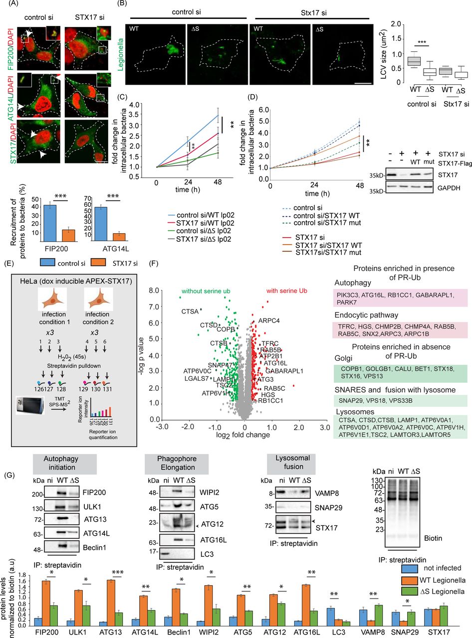
Figure Lengend Snippet: a) A549 cells were treated with control or STX17 siRNA for 48 h followed by infection with WT Legionella . Cells were fixed and stained for indicated autophagy markers 1 h after infection. The data are means ± SEM of 50 cells representing three experiments. which were analyzed per sample to quantitate recruitment of FIP200 and ATG14L to bacteria. p value was calculated using 2 tailed type 3 Student’s t-test, ***p=5.83E-7(FIP200), ***p=1.92E-12(ATG14L), Scale bar: 5µm. Dotted lines indicate cell outlines drawn from thresholding images in FIJI. b) A549 cells were treated with STX17 or control siRNA for 48 h followed by infection with WT or ΔS Legionella for 12 h (MOI = 1). Cells were fixed for immunostaining with a Legionella -specific antibody followed by confocal microscopy. The LCV size was estimated in FIJI. In the box-plot, center lines show the medians; box limits indicate the 25th and 75th percentiles as determined by R software; whiskers extend 1.5 times the interquartile range from the 25th and 75th percentiles, outliers are represented by dots. n = 32, 31 cells taken from 3 independent experiments. p value was calculated using 2 tailed, type 3 Student’s t-test, ***p =8.13E-16. Scale bar: 5µm. Dotted lines indicate cell outlines drawn from thresholding images in FIJI. c) A549 cells were treated with control or STX17 siRNA for 48 h followed by infection with WT or ΔS Legionella . Intracellular bacterial replication was assessed after 0, 24 and 48 h. Data are means ± SEM of three independent experiments. p value was calculated using 2 tailed type 3 Student’s t-test, **p =0.00526 (WT, control vs STX17siRNA, 24h), **p =0.00815 (WT, control vs STX17siRNA, 48h), d) A549 cells were treated with STX17 or control siRNA for 48 h followed by transfection with WT or PR-Ub-deficient STX17 for 24 h. Intracellular bacterial replication was assessed after 0, 24 and 48 h. Data are means ± SEM of three independent experiments **p=0.0077 (WT, STX17siRNA versus STX17 mutant, STX17siRNA, 48 h).Western blotting with STX17 antibody shows knockdown efficiency of STX17 siRNA and reconstitution with WT or mutant STX17. e) Proximity labeling assay workflow. HeLa cells expressing doxycycline-inducible APEX-STX17 and CD32(for increasing efficiency of Legionella uptake) were infected with Legionella for 2 h before treatment with biotin-tyramide and H 2 O 2 followed by streptavidin pulldown. The samples were reduced, alkylated and digested with trypsin before MS analysis. Samples representing three biological replicates each of non-infected and Legionella -infected cells were analyzed in a single reaction by 6plex TMT labeling. f) Volcano plot showing changes in the biotin-labeled proteome following the infection of HeLa cells expressing APEX2-FLAG-STX17 with ΔR and ΔRΔS Legionella for 2 h, GO analysis of the biotin-labeled proteome showing pathways upregulated by infection with ΔR vs ΔRΔS Legionella . Data represents mean fold change of three experimental replicates per infection set (n=3). p value was calculated using 2 tailed type 3 Student’s t-test and significant candidates were chosen having p-value ≤0.01 and log2(fold change) value minimum of ±0.5. Red and green indicate compartments containing proteins enriched following infection with ΔR and ΔRΔS Legionella , respectively. g) Cells expressing doxycycline-inducible APEX-STX17 were infected with Legionella for 2 h before treatment with biotin-tyramide and H 2 O 2 followed by streptavidin pulldown. The samples were analyzed by western blot with antibodies against proteins of the autophagic and endosomal pathways. The data represents means ± SD of 3 independent experiments. p value was calculated using 2 tailed type 3 Students t-test. WT vs ΔS p values: *p= 0.01006 (FIP200), **p=0.0206 (ULK1), p=***0.0007 (ATG13) **p=0.005 (ATG14), *p=0.0111(Beclin1), *p=0.0219 (WIPI2), **p=0.0038 (ATG5), *p=0.018 (ATG12), **p=0.001 (ATG16), **p=0.0036 (VAMP8), *p=0.0424 (SNAP29). (n.i-not infected, WT-wild-type Legionella , ΔS-ΔSidE Legionella )

Article Snippet: We used the following antibodies and dilutions: STX17 (cat. no. 17815-1-AP, Proteintech; 1:1000), GAPDH (cat. no. 2118, Cell Signaling Technology; 1:2000), GFP trap beads (cat. no. gta-100, ChromoTek), GFP (cat. no. sc-9996, Santa Cruz Biotechnology; 1:2000), GFP for immune-electron microscopy (cat no. 600–106-215, Rockland), biotin for immuno-electron microscopy (cat. no. 100–4198, Rockland), LC3 (cat. no. 2775, Cell Signaling Technology; 1:2000), VAMP8 (cat. no. 13060, Cell Signaling Technology; 1:1000), SNAP29 (cat. no. 3013, Cell Signaling Technology; 1:2000), RAB5 (cat. no. 3547; 1:1,000), RAB7 (cat. no. 9367, Cell Signaling Technology; 1:2000), ATG16L (cat. no. ab188642, Cell Signaling Technology; 1:1000), ATG12 (cat. no. 4180, Cell Signaling Technology; 1:1000), Beclin1 (cat. no. 3738, Cell Signaling Technology; 1:1000), FIP200 (cat. no. 17250-1AP, Proteintech; 1:1000), LAMP1 (cat. no. 9091, Cell Signaling Technology; 1:2000), and Legionella (cat. no. 20943, Abcam; 1:4000), Ubiquitin (Cat. no: 3933, Cell Signaling Technology, 1:1000).

Techniques: Control, Infection, Staining, Bacteria, Immunostaining, Confocal Microscopy, Software, Transfection, Mutagenesis, Western Blot, Knockdown, Labeling, Expressing