Review


pires puro3 plasmid  (TaKaRa)


Bioz Verified Symbol TaKaRa is a verified supplier
Bioz Manufacturer Symbol TaKaRa manufactures this product  
  • Logo
  • About
  • News
  • Press Release
  • Team
  • Advisors
  • Partners
  • Contact
  • Bioz Stars
  • Bioz vStars
  • 95

    Structured Review

    TaKaRa pires puro3 plasmid
    Pires Puro3 Plasmid, supplied by TaKaRa, used in various techniques. Bioz Stars score: 95/100, based on 373 PubMed citations. ZERO BIAS - scores, article reviews, protocol conditions and more
    https://www.bioz.com/product/pires+plasmid/pIRESpuro3+Vector/10__1038_slash_s41598___026___71371___y-52-15-17
    Average 95 stars, based on 373 article reviews
    pires puro3 plasmid - by Bioz Stars, 2026-11
    95/100 stars

    Images

    Related Articles

    Plasmid Preparation:

    Article Title: Virus-like particles which can be used in the treatment of autoimmune diseases
    Article Snippet: .. The plasmid pCTLA-4WT comprises a coding sequence for the wild-type CTLA-4 protein (NCBI Reference Sequence: nm_009843.4) inserted into the pIRES plasmid (Clontech®) using the EcoRI restriction site (position 1102). ..

    Article Title: Preferential expression of SCN1A in GABAergic neurons improves survival and epileptic phenotype in a mouse model of Dravet syndrome
    Article Snippet: Fragments from the human SCN1A 5′ UTR were amplified by PCR from genomic DNA (Table ), except for a 14-Kb fragment (F8) obtained from the BAC clone RP11-807A15 (Thermo Fisher Scientific). .. An intervening sequence (IVS) from the pIRES plasmid (Clontech) was placed between the SCN1A -based promoters and the firefly luciferase coding sequence in order to increase mRNA stability. .. Assembly of different combinations of sequences was performed using standard molecular biology techniques using restriction endonucleases from New England Biolabs.

    Article Title: Methods of propagating rhinovirus C in previously unsusceptible cell lines
    Article Snippet: .. The resulting amplicons were cloned into pIRES plasmid (Clontech) using restriction enzyme sites XhoI and XmaI to express the genes of interest under control of the CMV promoter. ..

    Article Title: Globally defining the effects of mutations in a picornavirus capsid
    Article Snippet: .. To test whether proteins were cleaved by the viral 3 CD protease, the corresponding region was PCR amplified from the Nancy infectious clone (primers 3C-For: TATTCTCGAGACCATGGGCCCTGCCTTTGAGTTCG and 3D-Rev: TATTGCGGCCGCCTAGAAGGAGTCCAACCATTTCCT ) and cloned into the pIRES plasmid (Clonetech) using the restriction sites XhoI and NotI (pIRES-3CD pro ). .. For analysis of fusion proteins, HEK293 cells were transfected with GFP-PLEKHA4 (kind gift of Dr. Jeremy Baskin, Cornell University), GFP-PLSCR1 (kind gift of Dr. Serge Benichou, Institut Cochin), pAcGFP-WDR33 (Kind gift of Dr. Matthias Altmeyer, University of Zurich), FLAG-NLCR5 (Addgene #37521), HA-ZC3HAV1 (Addgene #45907), or the control plasmid FLuc-eGFP (Addgene #90170), together with the pIRES-3CD pro plasmid using Lipofectamine 2000.

    Article Title: Human endothelin receptor antibody and use thereof
    Article Snippet: .. After 24 h culture, the cells were transfected with a pIRES plasmid (Clontech, commercial) modified to carry hETAR gene (see SEQ ID NO: 1 for the nucleotide sequence, and SEQ ID NO: 2 for the amino acid sequence). .. The medium was changed before transfection and the transfection was carried out by following the transfection conditions recommended by Invitrogen for Lipofectamine 2000.

    Article Title: Compositions and methods for self-regulated inducible gene expression
    Article Snippet: The arabinose expression plasmid is pBAD-DEST-49 (Invitrogen) and expression genes were placed within the first multiple cloning site. .. Eukaryotic Plasmids The pIRES plasmid (Clontech) has a minimal CMVI promoter with two multiple cloning sites linked by the IRES sequence. ..

    Sequencing:

    Article Title: Virus-like particles which can be used in the treatment of autoimmune diseases
    Article Snippet: .. The plasmid pCTLA-4WT comprises a coding sequence for the wild-type CTLA-4 protein (NCBI Reference Sequence: nm_009843.4) inserted into the pIRES plasmid (Clontech®) using the EcoRI restriction site (position 1102). ..

    Article Title: Preferential expression of SCN1A in GABAergic neurons improves survival and epileptic phenotype in a mouse model of Dravet syndrome
    Article Snippet: Fragments from the human SCN1A 5′ UTR were amplified by PCR from genomic DNA (Table ), except for a 14-Kb fragment (F8) obtained from the BAC clone RP11-807A15 (Thermo Fisher Scientific). .. An intervening sequence (IVS) from the pIRES plasmid (Clontech) was placed between the SCN1A -based promoters and the firefly luciferase coding sequence in order to increase mRNA stability. .. Assembly of different combinations of sequences was performed using standard molecular biology techniques using restriction endonucleases from New England Biolabs.

    Article Title: Human endothelin receptor antibody and use thereof
    Article Snippet: .. After 24 h culture, the cells were transfected with a pIRES plasmid (Clontech, commercial) modified to carry hETAR gene (see SEQ ID NO: 1 for the nucleotide sequence, and SEQ ID NO: 2 for the amino acid sequence). .. The medium was changed before transfection and the transfection was carried out by following the transfection conditions recommended by Invitrogen for Lipofectamine 2000.

    Article Title: Compositions and methods for self-regulated inducible gene expression
    Article Snippet: The arabinose expression plasmid is pBAD-DEST-49 (Invitrogen) and expression genes were placed within the first multiple cloning site. .. Eukaryotic Plasmids The pIRES plasmid (Clontech) has a minimal CMVI promoter with two multiple cloning sites linked by the IRES sequence. ..

    Luciferase:

    Article Title: Preferential expression of SCN1A in GABAergic neurons improves survival and epileptic phenotype in a mouse model of Dravet syndrome
    Article Snippet: Fragments from the human SCN1A 5′ UTR were amplified by PCR from genomic DNA (Table ), except for a 14-Kb fragment (F8) obtained from the BAC clone RP11-807A15 (Thermo Fisher Scientific). .. An intervening sequence (IVS) from the pIRES plasmid (Clontech) was placed between the SCN1A -based promoters and the firefly luciferase coding sequence in order to increase mRNA stability. .. Assembly of different combinations of sequences was performed using standard molecular biology techniques using restriction endonucleases from New England Biolabs.

    Clone Assay:

    Article Title: Methods of propagating rhinovirus C in previously unsusceptible cell lines
    Article Snippet: .. The resulting amplicons were cloned into pIRES plasmid (Clontech) using restriction enzyme sites XhoI and XmaI to express the genes of interest under control of the CMV promoter. ..

    Article Title: Globally defining the effects of mutations in a picornavirus capsid
    Article Snippet: .. To test whether proteins were cleaved by the viral 3 CD protease, the corresponding region was PCR amplified from the Nancy infectious clone (primers 3C-For: TATTCTCGAGACCATGGGCCCTGCCTTTGAGTTCG and 3D-Rev: TATTGCGGCCGCCTAGAAGGAGTCCAACCATTTCCT ) and cloned into the pIRES plasmid (Clonetech) using the restriction sites XhoI and NotI (pIRES-3CD pro ). .. For analysis of fusion proteins, HEK293 cells were transfected with GFP-PLEKHA4 (kind gift of Dr. Jeremy Baskin, Cornell University), GFP-PLSCR1 (kind gift of Dr. Serge Benichou, Institut Cochin), pAcGFP-WDR33 (Kind gift of Dr. Matthias Altmeyer, University of Zurich), FLAG-NLCR5 (Addgene #37521), HA-ZC3HAV1 (Addgene #45907), or the control plasmid FLuc-eGFP (Addgene #90170), together with the pIRES-3CD pro plasmid using Lipofectamine 2000.

    Control:

    Article Title: Methods of propagating rhinovirus C in previously unsusceptible cell lines
    Article Snippet: .. The resulting amplicons were cloned into pIRES plasmid (Clontech) using restriction enzyme sites XhoI and XmaI to express the genes of interest under control of the CMV promoter. ..

    Polymerase Chain Reaction:

    Article Title: Globally defining the effects of mutations in a picornavirus capsid
    Article Snippet: .. To test whether proteins were cleaved by the viral 3 CD protease, the corresponding region was PCR amplified from the Nancy infectious clone (primers 3C-For: TATTCTCGAGACCATGGGCCCTGCCTTTGAGTTCG and 3D-Rev: TATTGCGGCCGCCTAGAAGGAGTCCAACCATTTCCT ) and cloned into the pIRES plasmid (Clonetech) using the restriction sites XhoI and NotI (pIRES-3CD pro ). .. For analysis of fusion proteins, HEK293 cells were transfected with GFP-PLEKHA4 (kind gift of Dr. Jeremy Baskin, Cornell University), GFP-PLSCR1 (kind gift of Dr. Serge Benichou, Institut Cochin), pAcGFP-WDR33 (Kind gift of Dr. Matthias Altmeyer, University of Zurich), FLAG-NLCR5 (Addgene #37521), HA-ZC3HAV1 (Addgene #45907), or the control plasmid FLuc-eGFP (Addgene #90170), together with the pIRES-3CD pro plasmid using Lipofectamine 2000.

    Amplification:

    Article Title: Globally defining the effects of mutations in a picornavirus capsid
    Article Snippet: .. To test whether proteins were cleaved by the viral 3 CD protease, the corresponding region was PCR amplified from the Nancy infectious clone (primers 3C-For: TATTCTCGAGACCATGGGCCCTGCCTTTGAGTTCG and 3D-Rev: TATTGCGGCCGCCTAGAAGGAGTCCAACCATTTCCT ) and cloned into the pIRES plasmid (Clonetech) using the restriction sites XhoI and NotI (pIRES-3CD pro ). .. For analysis of fusion proteins, HEK293 cells were transfected with GFP-PLEKHA4 (kind gift of Dr. Jeremy Baskin, Cornell University), GFP-PLSCR1 (kind gift of Dr. Serge Benichou, Institut Cochin), pAcGFP-WDR33 (Kind gift of Dr. Matthias Altmeyer, University of Zurich), FLAG-NLCR5 (Addgene #37521), HA-ZC3HAV1 (Addgene #45907), or the control plasmid FLuc-eGFP (Addgene #90170), together with the pIRES-3CD pro plasmid using Lipofectamine 2000.

    Transfection:

    Article Title: Human endothelin receptor antibody and use thereof
    Article Snippet: .. After 24 h culture, the cells were transfected with a pIRES plasmid (Clontech, commercial) modified to carry hETAR gene (see SEQ ID NO: 1 for the nucleotide sequence, and SEQ ID NO: 2 for the amino acid sequence). .. The medium was changed before transfection and the transfection was carried out by following the transfection conditions recommended by Invitrogen for Lipofectamine 2000.

    Modification:

    Article Title: Human endothelin receptor antibody and use thereof
    Article Snippet: .. After 24 h culture, the cells were transfected with a pIRES plasmid (Clontech, commercial) modified to carry hETAR gene (see SEQ ID NO: 1 for the nucleotide sequence, and SEQ ID NO: 2 for the amino acid sequence). .. The medium was changed before transfection and the transfection was carried out by following the transfection conditions recommended by Invitrogen for Lipofectamine 2000.

    Cloning:

    Article Title: Compositions and methods for self-regulated inducible gene expression
    Article Snippet: The arabinose expression plasmid is pBAD-DEST-49 (Invitrogen) and expression genes were placed within the first multiple cloning site. .. Eukaryotic Plasmids The pIRES plasmid (Clontech) has a minimal CMVI promoter with two multiple cloning sites linked by the IRES sequence. ..


    Similar Products

    95
    TaKaRa pires puro3 plasmid
    Pires Puro3 Plasmid, supplied by TaKaRa, used in various techniques. Bioz Stars score: 95/100, based on 1 PubMed citations. ZERO BIAS - scores, article reviews, protocol conditions and more
    https://www.bioz.com/product/pires+plasmid/pIRESpuro3+Vector/10__1038_slash_s41598___026___71371___y-52-15-17
    Average 95 stars, based on 1 article reviews
    pires puro3 plasmid - by Bioz Stars, 2026-11
    95/100 stars
      Buy from Supplier

    93
    Addgene inc pires egfp pac
    Pires Egfp Pac, supplied by Addgene inc, used in various techniques. Bioz Stars score: 93/100, based on 1 PubMed citations. ZERO BIAS - scores, article reviews, protocol conditions and more
    https://www.bioz.com/product/pires+plasmid/pIRES-EGFP-puro+(Plasmid+%2345567)/pmc12825063-47-0-2
    Average 93 stars, based on 1 article reviews
    pires egfp pac - by Bioz Stars, 2026-11
    93/100 stars
      Buy from Supplier

    93
    Addgene inc flag addgene 16257 pires neo3 clontech 631621 aspn d14
    Flag Addgene 16257 Pires Neo3 Clontech 631621 Aspn D14, supplied by Addgene inc, used in various techniques. Bioz Stars score: 93/100, based on 1 PubMed citations. ZERO BIAS - scores, article reviews, protocol conditions and more
    https://www.bioz.com/product/pires+plasmid/HER2+WT+(Plasmid+%2316257)/pmc12406728__jciinsight-10-187151-s057-88-282-283
    Average 93 stars, based on 1 article reviews
    flag addgene 16257 pires neo3 clontech 631621 aspn d14 - by Bioz Stars, 2026-11
    93/100 stars
      Buy from Supplier

    93
    Addgene inc pires 2 egfp vector
    Pires 2 Egfp Vector, supplied by Addgene inc, used in various techniques. Bioz Stars score: 93/100, based on 1 PubMed citations. ZERO BIAS - scores, article reviews, protocol conditions and more
    https://www.bioz.com/product/pires+plasmid/pIRES2-EGFP+Mer+(Plasmid+%2314998)/bio_rxiv__2025__07__16__665058-25-17-21
    Average 93 stars, based on 1 article reviews
    pires 2 egfp vector - by Bioz Stars, 2026-11
    93/100 stars
      Buy from Supplier

    92
    Addgene inc pires neo flag ha eyfp
    ( A ) Schematic overview of the structure of pUS28. All structural parts of the protein and the DRY motif are indicated. Mutation of the arginine in this motif to alanine (R129A) ablates G-protein coupling. This panel was created using BioRender.com . ( B ) HeLa cells were either left untreated or were co-transfected with CIITA-3xFLAG expression construct and pcDNA:US28-HA, pcDNA:US28-R129A-HA, pIRESNeo-FLAG/HA-EYFP, or empty vector. At 24 hr post-transfection, protein lysates were generated and analyzed by immunoblot using antibodies detecting the indicated proteins or the respective epitope tags. ( C ) HeLa cells were either left untreated or were co-transfected with CIITA-3xFLAG expression construct and pcDNA:US28-HA, pcDNA:US28-ΔC-HA, pIRESNeo-FLAG/HA-EYFP (control) or empty vector. At 24 hr post-transfection, protein lysates were generated and analyzed by immunoblot using antibodies detecting the indicated proteins or the respective epitope tags. ( D/E ) HeLa cells were either left untreated or were co-transfected with CIITA expression construct and pcDNA:US28-HA, pcDNA:US28-R129A-HA, pIRESNeo-FLAG/HA-EYFP (control) or empty vector. At 48 hr post-transfection, cells were stained with anti-HLA-DP ( D ) or anti-HLA-DR ( E ) antibodies and analyzed by flow cytometry. Cell surface expression of HLA-DP or HLA-DR was normalized to cells co-transfected with CIITA expression construct and empty vector. The percentage of HLA-DP- and HLA-DR-positive cells is shown (n=3). The different transfection conditions were compared to the control condition (vector) by one-way ANOVA test. Additionally, US28-R129A-HA was compared to US28-HA. Comparisons are shown when statistically significant. ( F/G ) HeLa cells were either left untreated or were co-transfected with CIITA-3xFLAG expression construct and pcDNA:US28-HA, pcDNA:US28-ΔC-HA, pIRESNeo-FLAG/HA-EYFP (control) or empty vector. At 48 hr post-transfection, cells were stained with anti-HLA-DP ( F ) or anti-HLA-DR ( G ) antibodies and analyzed by flow cytometry. Cell surface expression of HLA-DP or HLA-DR was normalized to cells transfected with CIITA expression construct and empty vector. The percentage of HLA-DP- and HLA-DR-positive cells is shown (n=3). The different transfection conditions were compared to the control condition (vector) by one-way ANOVA test. Additionally, US28-ΔC-HA was compared to US28-HA. Comparisons are shown when statistically significant. ( H ) HeLa cells were either left untreated or were transfected with CIITA-3xFLAG expression construct or pcDNA:US28-ΔC-HA. At 24 hr post-transfection, protein lysates were generated and an IP with HA-specific mouse monoclonal antibody was performed either with unmodified samples or with mixed samples of CIITA- and US28-transfected cells. The lysates and IP samples were analyzed by immunoblot to test CIITA and pUS28-ΔC co-precipitation. ( I ) HeLa cells were either left untreated or were co-transfected with CIITA-3xFLAG expression construct and <t>pIRES:US28-HA,</t> different pIRES:US28-ΔC-HA mutants, pIRESNeo-FLAG/HA-EYFP (control) or empty vector. At 24 hr post-transfection, protein lysates were generated and analyzed by immunoblot using antibodies detecting the indicated proteins or the respective epitope tags. Untrans, untransfected. Vector, empty vector control. ΔC (1-297), C-terminal (aa 298–354) deletion mutant of pUS28. 1–313, C-terminal (aa 314–354) deletion mutant of pUS28. 1–329, C-terminal (aa 330–354) deletion mutant of pUS28. 1–345, C-terminal (aa 346–354) deletion mutant of pUS28. R129A, point mutation mutant of pUS28. Control, pIRESNeo-FLAG/HA-EYFP. Figure 7—source data 1. PDF files containing original western blots for , indicating the relevant bands and treatments. Figure 7—source data 2. Original files for western blot analysis displayed in .
    Pires Neo Flag Ha Eyfp, supplied by Addgene inc, used in various techniques. Bioz Stars score: 92/100, based on 1 PubMed citations. ZERO BIAS - scores, article reviews, protocol conditions and more
    https://www.bioz.com/product/pires+plasmid/pIRESneo-FLAG%2FHA+EYFP+(Plasmid+%2310825)/pmc12226020-200-15-19
    Average 92 stars, based on 1 article reviews
    pires neo flag ha eyfp - by Bioz Stars, 2026-11
    92/100 stars
      Buy from Supplier

    93
    Addgene inc michael mcvoy
    ( A ) Schematic overview of the structure of pUS28. All structural parts of the protein and the DRY motif are indicated. Mutation of the arginine in this motif to alanine (R129A) ablates G-protein coupling. This panel was created using BioRender.com . ( B ) HeLa cells were either left untreated or were co-transfected with CIITA-3xFLAG expression construct and pcDNA:US28-HA, pcDNA:US28-R129A-HA, pIRESNeo-FLAG/HA-EYFP, or empty vector. At 24 hr post-transfection, protein lysates were generated and analyzed by immunoblot using antibodies detecting the indicated proteins or the respective epitope tags. ( C ) HeLa cells were either left untreated or were co-transfected with CIITA-3xFLAG expression construct and pcDNA:US28-HA, pcDNA:US28-ΔC-HA, pIRESNeo-FLAG/HA-EYFP (control) or empty vector. At 24 hr post-transfection, protein lysates were generated and analyzed by immunoblot using antibodies detecting the indicated proteins or the respective epitope tags. ( D/E ) HeLa cells were either left untreated or were co-transfected with CIITA expression construct and pcDNA:US28-HA, pcDNA:US28-R129A-HA, pIRESNeo-FLAG/HA-EYFP (control) or empty vector. At 48 hr post-transfection, cells were stained with anti-HLA-DP ( D ) or anti-HLA-DR ( E ) antibodies and analyzed by flow cytometry. Cell surface expression of HLA-DP or HLA-DR was normalized to cells co-transfected with CIITA expression construct and empty vector. The percentage of HLA-DP- and HLA-DR-positive cells is shown (n=3). The different transfection conditions were compared to the control condition (vector) by one-way ANOVA test. Additionally, US28-R129A-HA was compared to US28-HA. Comparisons are shown when statistically significant. ( F/G ) HeLa cells were either left untreated or were co-transfected with CIITA-3xFLAG expression construct and pcDNA:US28-HA, pcDNA:US28-ΔC-HA, pIRESNeo-FLAG/HA-EYFP (control) or empty vector. At 48 hr post-transfection, cells were stained with anti-HLA-DP ( F ) or anti-HLA-DR ( G ) antibodies and analyzed by flow cytometry. Cell surface expression of HLA-DP or HLA-DR was normalized to cells transfected with CIITA expression construct and empty vector. The percentage of HLA-DP- and HLA-DR-positive cells is shown (n=3). The different transfection conditions were compared to the control condition (vector) by one-way ANOVA test. Additionally, US28-ΔC-HA was compared to US28-HA. Comparisons are shown when statistically significant. ( H ) HeLa cells were either left untreated or were transfected with CIITA-3xFLAG expression construct or pcDNA:US28-ΔC-HA. At 24 hr post-transfection, protein lysates were generated and an IP with HA-specific mouse monoclonal antibody was performed either with unmodified samples or with mixed samples of CIITA- and US28-transfected cells. The lysates and IP samples were analyzed by immunoblot to test CIITA and pUS28-ΔC co-precipitation. ( I ) HeLa cells were either left untreated or were co-transfected with CIITA-3xFLAG expression construct and <t>pIRES:US28-HA,</t> different pIRES:US28-ΔC-HA mutants, pIRESNeo-FLAG/HA-EYFP (control) or empty vector. At 24 hr post-transfection, protein lysates were generated and analyzed by immunoblot using antibodies detecting the indicated proteins or the respective epitope tags. Untrans, untransfected. Vector, empty vector control. ΔC (1-297), C-terminal (aa 298–354) deletion mutant of pUS28. 1–313, C-terminal (aa 314–354) deletion mutant of pUS28. 1–329, C-terminal (aa 330–354) deletion mutant of pUS28. 1–345, C-terminal (aa 346–354) deletion mutant of pUS28. R129A, point mutation mutant of pUS28. Control, pIRESNeo-FLAG/HA-EYFP. Figure 7—source data 1. PDF files containing original western blots for , indicating the relevant bands and treatments. Figure 7—source data 2. Original files for western blot analysis displayed in .
    Michael Mcvoy, supplied by Addgene inc, used in various techniques. Bioz Stars score: 93/100, based on 1 PubMed citations. ZERO BIAS - scores, article reviews, protocol conditions and more
    https://www.bioz.com/product/pires+plasmid/pIRES-EGFP-puro+(Plasmid+%2345567)/pm40533368-212-29-31
    Average 93 stars, based on 1 article reviews
    michael mcvoy - by Bioz Stars, 2026-11
    93/100 stars
      Buy from Supplier

    93
    Addgene inc mammalian expression vector pires cyofp1
    Averaged whole cell current traces from cells expressing KCNE1 variants in the homozygous state. Each trace represents JNJ-303 sensitive currents from an average of 21-126 cells co-expressing <t>KCNQ1</t> with the indicated KCNE1 variant. Currents were normalized to peak current in cells expressing WT KCNE1 and WT KCNQ1 recorded in parallel. Scale bars represent 25% of WT (vertical) and 500 ms (horizontal). Location of variants is indicated by the colored labels (green = N-terminus; orange = TM domain; blue = C-terminus).
    Mammalian Expression Vector Pires Cyofp1, supplied by Addgene inc, used in various techniques. Bioz Stars score: 93/100, based on 1 PubMed citations. ZERO BIAS - scores, article reviews, protocol conditions and more
    https://www.bioz.com/product/pires+plasmid/NHR1%2B2-Flag+pcDNA3%2E1+(Plasmid+%2317316)/bio_rxiv__2025__03__28__646046-36-15-20
    Average 93 stars, based on 1 article reviews
    mammalian expression vector pires cyofp1 - by Bioz Stars, 2026-11
    93/100 stars
      Buy from Supplier

    93
    Addgene inc pires hrgfp ii tet1 cd
    Averaged whole cell current traces from cells expressing KCNE1 variants in the homozygous state. Each trace represents JNJ-303 sensitive currents from an average of 21-126 cells co-expressing <t>KCNQ1</t> with the indicated KCNE1 variant. Currents were normalized to peak current in cells expressing WT KCNE1 and WT KCNQ1 recorded in parallel. Scale bars represent 25% of WT (vertical) and 500 ms (horizontal). Location of variants is indicated by the colored labels (green = N-terminus; orange = TM domain; blue = C-terminus).
    Pires Hrgfp Ii Tet1 Cd, supplied by Addgene inc, used in various techniques. Bioz Stars score: 93/100, based on 1 PubMed citations. ZERO BIAS - scores, article reviews, protocol conditions and more
    https://www.bioz.com/product/pires+plasmid/pIRES-hrGFP+II-TET1_CD+(Plasmid+%2383570)/pmc12081649-16-0-3
    Average 93 stars, based on 1 article reviews
    pires hrgfp ii tet1 cd - by Bioz Stars, 2026-11
    93/100 stars
      Buy from Supplier

    Image Search Results


    ( A ) Schematic overview of the structure of pUS28. All structural parts of the protein and the DRY motif are indicated. Mutation of the arginine in this motif to alanine (R129A) ablates G-protein coupling. This panel was created using BioRender.com . ( B ) HeLa cells were either left untreated or were co-transfected with CIITA-3xFLAG expression construct and pcDNA:US28-HA, pcDNA:US28-R129A-HA, pIRESNeo-FLAG/HA-EYFP, or empty vector. At 24 hr post-transfection, protein lysates were generated and analyzed by immunoblot using antibodies detecting the indicated proteins or the respective epitope tags. ( C ) HeLa cells were either left untreated or were co-transfected with CIITA-3xFLAG expression construct and pcDNA:US28-HA, pcDNA:US28-ΔC-HA, pIRESNeo-FLAG/HA-EYFP (control) or empty vector. At 24 hr post-transfection, protein lysates were generated and analyzed by immunoblot using antibodies detecting the indicated proteins or the respective epitope tags. ( D/E ) HeLa cells were either left untreated or were co-transfected with CIITA expression construct and pcDNA:US28-HA, pcDNA:US28-R129A-HA, pIRESNeo-FLAG/HA-EYFP (control) or empty vector. At 48 hr post-transfection, cells were stained with anti-HLA-DP ( D ) or anti-HLA-DR ( E ) antibodies and analyzed by flow cytometry. Cell surface expression of HLA-DP or HLA-DR was normalized to cells co-transfected with CIITA expression construct and empty vector. The percentage of HLA-DP- and HLA-DR-positive cells is shown (n=3). The different transfection conditions were compared to the control condition (vector) by one-way ANOVA test. Additionally, US28-R129A-HA was compared to US28-HA. Comparisons are shown when statistically significant. ( F/G ) HeLa cells were either left untreated or were co-transfected with CIITA-3xFLAG expression construct and pcDNA:US28-HA, pcDNA:US28-ΔC-HA, pIRESNeo-FLAG/HA-EYFP (control) or empty vector. At 48 hr post-transfection, cells were stained with anti-HLA-DP ( F ) or anti-HLA-DR ( G ) antibodies and analyzed by flow cytometry. Cell surface expression of HLA-DP or HLA-DR was normalized to cells transfected with CIITA expression construct and empty vector. The percentage of HLA-DP- and HLA-DR-positive cells is shown (n=3). The different transfection conditions were compared to the control condition (vector) by one-way ANOVA test. Additionally, US28-ΔC-HA was compared to US28-HA. Comparisons are shown when statistically significant. ( H ) HeLa cells were either left untreated or were transfected with CIITA-3xFLAG expression construct or pcDNA:US28-ΔC-HA. At 24 hr post-transfection, protein lysates were generated and an IP with HA-specific mouse monoclonal antibody was performed either with unmodified samples or with mixed samples of CIITA- and US28-transfected cells. The lysates and IP samples were analyzed by immunoblot to test CIITA and pUS28-ΔC co-precipitation. ( I ) HeLa cells were either left untreated or were co-transfected with CIITA-3xFLAG expression construct and pIRES:US28-HA, different pIRES:US28-ΔC-HA mutants, pIRESNeo-FLAG/HA-EYFP (control) or empty vector. At 24 hr post-transfection, protein lysates were generated and analyzed by immunoblot using antibodies detecting the indicated proteins or the respective epitope tags. Untrans, untransfected. Vector, empty vector control. ΔC (1-297), C-terminal (aa 298–354) deletion mutant of pUS28. 1–313, C-terminal (aa 314–354) deletion mutant of pUS28. 1–329, C-terminal (aa 330–354) deletion mutant of pUS28. 1–345, C-terminal (aa 346–354) deletion mutant of pUS28. R129A, point mutation mutant of pUS28. Control, pIRESNeo-FLAG/HA-EYFP. Figure 7—source data 1. PDF files containing original western blots for , indicating the relevant bands and treatments. Figure 7—source data 2. Original files for western blot analysis displayed in .

    Journal: eLife

    Article Title: The human cytomegalovirus-encoded pUS28 antagonizes CD4+ T cell recognition by targeting CIITA

    doi: 10.7554/eLife.96414

    Figure Lengend Snippet: ( A ) Schematic overview of the structure of pUS28. All structural parts of the protein and the DRY motif are indicated. Mutation of the arginine in this motif to alanine (R129A) ablates G-protein coupling. This panel was created using BioRender.com . ( B ) HeLa cells were either left untreated or were co-transfected with CIITA-3xFLAG expression construct and pcDNA:US28-HA, pcDNA:US28-R129A-HA, pIRESNeo-FLAG/HA-EYFP, or empty vector. At 24 hr post-transfection, protein lysates were generated and analyzed by immunoblot using antibodies detecting the indicated proteins or the respective epitope tags. ( C ) HeLa cells were either left untreated or were co-transfected with CIITA-3xFLAG expression construct and pcDNA:US28-HA, pcDNA:US28-ΔC-HA, pIRESNeo-FLAG/HA-EYFP (control) or empty vector. At 24 hr post-transfection, protein lysates were generated and analyzed by immunoblot using antibodies detecting the indicated proteins or the respective epitope tags. ( D/E ) HeLa cells were either left untreated or were co-transfected with CIITA expression construct and pcDNA:US28-HA, pcDNA:US28-R129A-HA, pIRESNeo-FLAG/HA-EYFP (control) or empty vector. At 48 hr post-transfection, cells were stained with anti-HLA-DP ( D ) or anti-HLA-DR ( E ) antibodies and analyzed by flow cytometry. Cell surface expression of HLA-DP or HLA-DR was normalized to cells co-transfected with CIITA expression construct and empty vector. The percentage of HLA-DP- and HLA-DR-positive cells is shown (n=3). The different transfection conditions were compared to the control condition (vector) by one-way ANOVA test. Additionally, US28-R129A-HA was compared to US28-HA. Comparisons are shown when statistically significant. ( F/G ) HeLa cells were either left untreated or were co-transfected with CIITA-3xFLAG expression construct and pcDNA:US28-HA, pcDNA:US28-ΔC-HA, pIRESNeo-FLAG/HA-EYFP (control) or empty vector. At 48 hr post-transfection, cells were stained with anti-HLA-DP ( F ) or anti-HLA-DR ( G ) antibodies and analyzed by flow cytometry. Cell surface expression of HLA-DP or HLA-DR was normalized to cells transfected with CIITA expression construct and empty vector. The percentage of HLA-DP- and HLA-DR-positive cells is shown (n=3). The different transfection conditions were compared to the control condition (vector) by one-way ANOVA test. Additionally, US28-ΔC-HA was compared to US28-HA. Comparisons are shown when statistically significant. ( H ) HeLa cells were either left untreated or were transfected with CIITA-3xFLAG expression construct or pcDNA:US28-ΔC-HA. At 24 hr post-transfection, protein lysates were generated and an IP with HA-specific mouse monoclonal antibody was performed either with unmodified samples or with mixed samples of CIITA- and US28-transfected cells. The lysates and IP samples were analyzed by immunoblot to test CIITA and pUS28-ΔC co-precipitation. ( I ) HeLa cells were either left untreated or were co-transfected with CIITA-3xFLAG expression construct and pIRES:US28-HA, different pIRES:US28-ΔC-HA mutants, pIRESNeo-FLAG/HA-EYFP (control) or empty vector. At 24 hr post-transfection, protein lysates were generated and analyzed by immunoblot using antibodies detecting the indicated proteins or the respective epitope tags. Untrans, untransfected. Vector, empty vector control. ΔC (1-297), C-terminal (aa 298–354) deletion mutant of pUS28. 1–313, C-terminal (aa 314–354) deletion mutant of pUS28. 1–329, C-terminal (aa 330–354) deletion mutant of pUS28. 1–345, C-terminal (aa 346–354) deletion mutant of pUS28. R129A, point mutation mutant of pUS28. Control, pIRESNeo-FLAG/HA-EYFP. Figure 7—source data 1. PDF files containing original western blots for , indicating the relevant bands and treatments. Figure 7—source data 2. Original files for western blot analysis displayed in .

    Article Snippet: Cells were transfected with the following plasmids: pUNO1-hCIITA (Invitrogen), pRP-humanCIITA-3xFLAG (VectorBuilder), pRP-3xFLAG-mouseCIITA (VectorBuilder), pcDNA3.1(+) (Invitrogen), pIRES neo -FLAG/HA-EYFP (RRID: Addgene_10825 , Gift from Thomas Tuschl ), pOG44 (Invitrogen), and HCMV ORF Expression Library ( ).

    Techniques: Mutagenesis, Transfection, Expressing, Construct, Plasmid Preparation, Generated, Western Blot, Control, Staining, Flow Cytometry

    Averaged whole cell current traces from cells expressing KCNE1 variants in the homozygous state. Each trace represents JNJ-303 sensitive currents from an average of 21-126 cells co-expressing KCNQ1 with the indicated KCNE1 variant. Currents were normalized to peak current in cells expressing WT KCNE1 and WT KCNQ1 recorded in parallel. Scale bars represent 25% of WT (vertical) and 500 ms (horizontal). Location of variants is indicated by the colored labels (green = N-terminus; orange = TM domain; blue = C-terminus).

    Journal: bioRxiv

    Article Title: Functional profiling of KCNE1 variants informs population carrier frequency of Jervell and Lange-Nielsen syndrome type 2

    doi: 10.1101/2025.03.28.646046

    Figure Lengend Snippet: Averaged whole cell current traces from cells expressing KCNE1 variants in the homozygous state. Each trace represents JNJ-303 sensitive currents from an average of 21-126 cells co-expressing KCNQ1 with the indicated KCNE1 variant. Currents were normalized to peak current in cells expressing WT KCNE1 and WT KCNQ1 recorded in parallel. Scale bars represent 25% of WT (vertical) and 500 ms (horizontal). Location of variants is indicated by the colored labels (green = N-terminus; orange = TM domain; blue = C-terminus).

    Article Snippet: Full length cDNA encoding WT KCNQ1 (GenBank accession number AF000571 ) was engineered in the mammalian expression vector pIRES-CyOFP1 (pIRES-CyOFP1-KCNQ1, AddGene #173162).

    Techniques: Expressing, Variant Assay

    Peak current density for KCNE1 variants in the homozygous state. JNJ-303 sensitive peak whole-cell currents measured at +60 mV from CHO-K1 cells co-expressing WT KCNQ1 with KCNE1 variants plotted as fold difference from WT channels recorded in parallel. ( A ) Variants in the N-terminus (green symbols). ( B ) Variants in the transmembrane domain (orange symbols). ( C ) Variants in the C-terminus (blue symbols). All individual data points are plotted as open symbols and mean values are shown as filled symbols with error bars representing the 95% CI. Values to the right or left of the vertical dashed line (normalized WT value) represent current density larger (gain-of-function) or smaller (loss-of-function) than WT, respectively. The number of recorded cells for each variant and individual P -values are presented in Supplemental Dataset 1 .

    Journal: bioRxiv

    Article Title: Functional profiling of KCNE1 variants informs population carrier frequency of Jervell and Lange-Nielsen syndrome type 2

    doi: 10.1101/2025.03.28.646046

    Figure Lengend Snippet: Peak current density for KCNE1 variants in the homozygous state. JNJ-303 sensitive peak whole-cell currents measured at +60 mV from CHO-K1 cells co-expressing WT KCNQ1 with KCNE1 variants plotted as fold difference from WT channels recorded in parallel. ( A ) Variants in the N-terminus (green symbols). ( B ) Variants in the transmembrane domain (orange symbols). ( C ) Variants in the C-terminus (blue symbols). All individual data points are plotted as open symbols and mean values are shown as filled symbols with error bars representing the 95% CI. Values to the right or left of the vertical dashed line (normalized WT value) represent current density larger (gain-of-function) or smaller (loss-of-function) than WT, respectively. The number of recorded cells for each variant and individual P -values are presented in Supplemental Dataset 1 .

    Article Snippet: Full length cDNA encoding WT KCNQ1 (GenBank accession number AF000571 ) was engineered in the mammalian expression vector pIRES-CyOFP1 (pIRES-CyOFP1-KCNQ1, AddGene #173162).

    Techniques: Expressing, Variant Assay

    Averaged whole cell current traces from cells expressing KCNE1 variants in the heterozygous state. Each trace represents JNJ-303 sensitive currents from an average of 38-131 CHO-E1 cells coexpressing KCNQ1 with the indicated KCNE1 variant. Currents were normalized to peak current in CHO-E1 cells co-expressing WT KCNE1 and WT KCNQ1 recorded in parallel. Scale bars represent 25% of WT (vertical) and 500 ms (horizontal). Location of variants is indicated by the colored labels (green = N-terminus; orange = TM domain; blue = C-terminus).

    Journal: bioRxiv

    Article Title: Functional profiling of KCNE1 variants informs population carrier frequency of Jervell and Lange-Nielsen syndrome type 2

    doi: 10.1101/2025.03.28.646046

    Figure Lengend Snippet: Averaged whole cell current traces from cells expressing KCNE1 variants in the heterozygous state. Each trace represents JNJ-303 sensitive currents from an average of 38-131 CHO-E1 cells coexpressing KCNQ1 with the indicated KCNE1 variant. Currents were normalized to peak current in CHO-E1 cells co-expressing WT KCNE1 and WT KCNQ1 recorded in parallel. Scale bars represent 25% of WT (vertical) and 500 ms (horizontal). Location of variants is indicated by the colored labels (green = N-terminus; orange = TM domain; blue = C-terminus).

    Article Snippet: Full length cDNA encoding WT KCNQ1 (GenBank accession number AF000571 ) was engineered in the mammalian expression vector pIRES-CyOFP1 (pIRES-CyOFP1-KCNQ1, AddGene #173162).

    Techniques: Expressing, Variant Assay

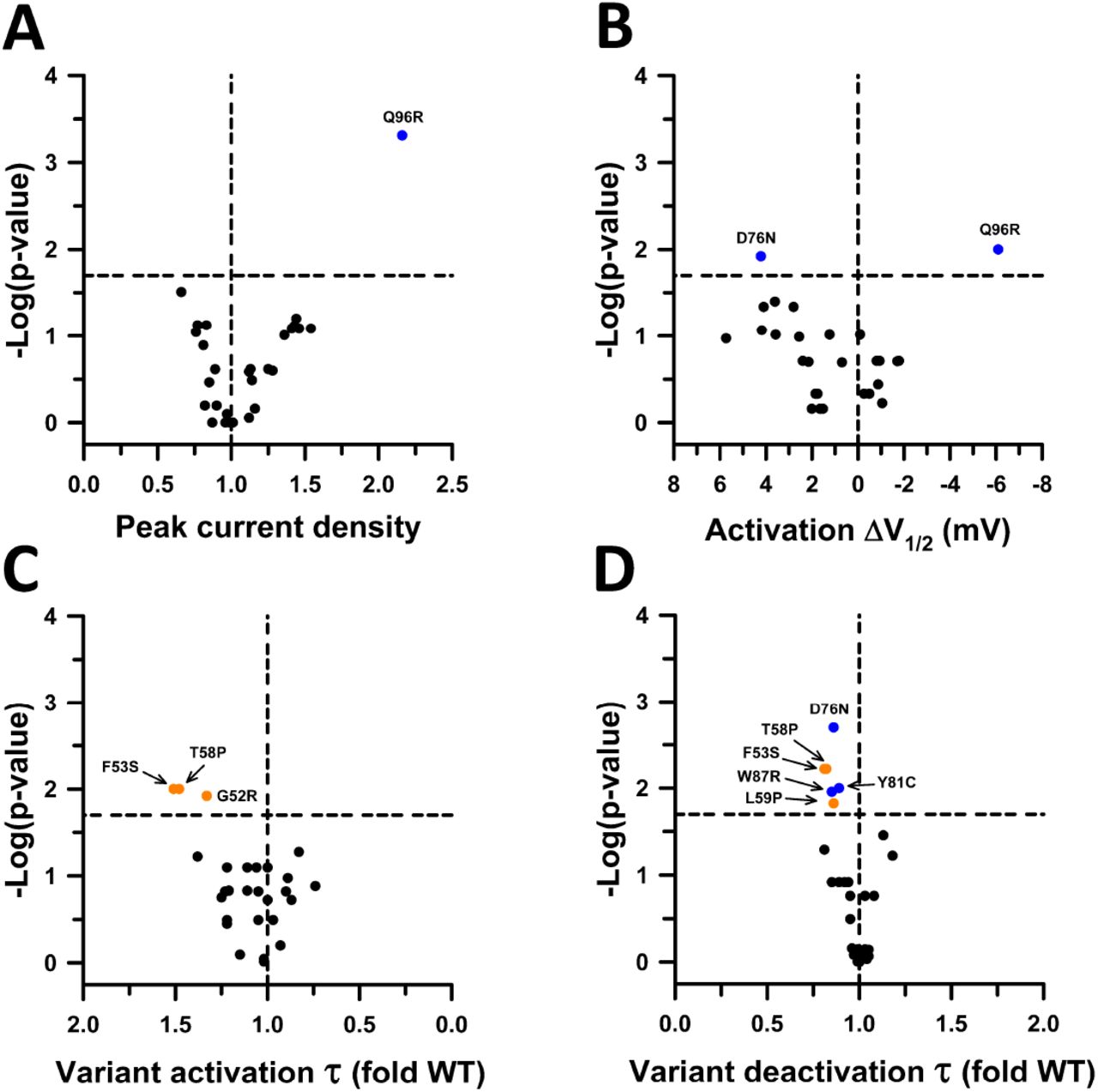
    Functional properties of KCNE1 variants in the heterozygous state. ( A ) Volcano plots of mean JNJ-303 sensitive peak whole-cell current density measured at +60 mV from CHO-E1 cells co-expressing WT KCNQ1 with each KCNE1 variant. Data are displayed as fold divergence from WT channels recorded in parallel. Only variants with peak current density significantly different from WT ( P < 0.02, horizontal dashed line) are labeled. Values to the right or left of the vertical dashed line (normalized WT value) represent current density larger or smaller than WT, respectively. ( B ) Volcano plots of mean difference (ΔV½ in mV) between the averaged V½ for variant and WT channels recorded in parallel. Only variants with peak current density significantly different from WT (P<0.02, horizontal dashed line) are labeled. Values to the right or left of the vertical dashed line indicate hyperpolarized or depolarized activation V½, respectively. ( C ) Volcano plots of mean activation time constants (τ) determined for each heterozygous KCNE1 variant plotted as the ratio to the averaged activation time constant for WT channels recorded in parallel. Values to the right or left of the vertical dashed line (no difference from WT) indicate faster or slower activation, respectively. ( D ) Volcano plots of mean deactivation time constants (τ) determined for each heterozygous KCNE1 variant plotted as the ratio to the averaged deactivation time constant for WT channels recorded in parallel. Values to the right or left of the vertical dashed line indicate slower or faster deactivation, respectively. Symbols colors are defined in previous figures. The number of recorded cells for each variant and individual P -values are presented in Supplemental Dataset 2 . Scatter plots of individual data are presented in Supplemental Figure S8 .

    Journal: bioRxiv

    Article Title: Functional profiling of KCNE1 variants informs population carrier frequency of Jervell and Lange-Nielsen syndrome type 2

    doi: 10.1101/2025.03.28.646046

    Figure Lengend Snippet: Functional properties of KCNE1 variants in the heterozygous state. ( A ) Volcano plots of mean JNJ-303 sensitive peak whole-cell current density measured at +60 mV from CHO-E1 cells co-expressing WT KCNQ1 with each KCNE1 variant. Data are displayed as fold divergence from WT channels recorded in parallel. Only variants with peak current density significantly different from WT ( P < 0.02, horizontal dashed line) are labeled. Values to the right or left of the vertical dashed line (normalized WT value) represent current density larger or smaller than WT, respectively. ( B ) Volcano plots of mean difference (ΔV½ in mV) between the averaged V½ for variant and WT channels recorded in parallel. Only variants with peak current density significantly different from WT (P<0.02, horizontal dashed line) are labeled. Values to the right or left of the vertical dashed line indicate hyperpolarized or depolarized activation V½, respectively. ( C ) Volcano plots of mean activation time constants (τ) determined for each heterozygous KCNE1 variant plotted as the ratio to the averaged activation time constant for WT channels recorded in parallel. Values to the right or left of the vertical dashed line (no difference from WT) indicate faster or slower activation, respectively. ( D ) Volcano plots of mean deactivation time constants (τ) determined for each heterozygous KCNE1 variant plotted as the ratio to the averaged deactivation time constant for WT channels recorded in parallel. Values to the right or left of the vertical dashed line indicate slower or faster deactivation, respectively. Symbols colors are defined in previous figures. The number of recorded cells for each variant and individual P -values are presented in Supplemental Dataset 2 . Scatter plots of individual data are presented in Supplemental Figure S8 .

    Article Snippet: Full length cDNA encoding WT KCNQ1 (GenBank accession number AF000571 ) was engineered in the mammalian expression vector pIRES-CyOFP1 (pIRES-CyOFP1-KCNQ1, AddGene #173162).

    Techniques: Functional Assay, Expressing, Variant Assay, Labeling, Activation Assay

    Functional properties of KCNE1 variants in the heterozygous state. ( A ) Average JNJ-303-sensitive peak whole-cell current density measured at +60 mV from CHO-E1 cells co-expressing WT KCNQ1 with each KCNE1 variant. Data are displayed as fold divergence from WT channels recorded in parallel. Values to the right or left of the vertical dashed line (normalized WT value) represent current density larger or smaller than WT, respectively. ( B ) Activation voltage-dependence for each heterozygous KCNE1 variant plotted as the difference (ΔV½ in mV) between the averaged V½ for WT channels recorded in parallel. Values to the right or left of the vertical dashed line indicate hyperpolarized or depolarized activation V½, respectively. ( C ) Activation time constants (τ) determined for each heterozygous KCNE1 variant plotted as the ratio to the averaged activation time constant for WT channels recorded in parallel. Values to the right or left of the vertical dashed line (no difference from WT) indicate faster or slower activation, respectively. ( D ) Deactivation time constants (τ) determined for each heterozygous KCNE1 variant plotted as the ratio to the averaged deactivation time constant for WT channels recorded in parallel. Values to the right or left of the vertical dashed line indicate slower or faster deactivation, respectively. Location of variants is indicated by the colored symbols (green = N-terminus; orange = TM domain; blue = C-terminus). The number of recorded cells for each variant and individual P -values are presented in Dataset 2 .

    Journal: bioRxiv

    Article Title: Functional profiling of KCNE1 variants informs population carrier frequency of Jervell and Lange-Nielsen syndrome type 2

    doi: 10.1101/2025.03.28.646046

    Figure Lengend Snippet: Functional properties of KCNE1 variants in the heterozygous state. ( A ) Average JNJ-303-sensitive peak whole-cell current density measured at +60 mV from CHO-E1 cells co-expressing WT KCNQ1 with each KCNE1 variant. Data are displayed as fold divergence from WT channels recorded in parallel. Values to the right or left of the vertical dashed line (normalized WT value) represent current density larger or smaller than WT, respectively. ( B ) Activation voltage-dependence for each heterozygous KCNE1 variant plotted as the difference (ΔV½ in mV) between the averaged V½ for WT channels recorded in parallel. Values to the right or left of the vertical dashed line indicate hyperpolarized or depolarized activation V½, respectively. ( C ) Activation time constants (τ) determined for each heterozygous KCNE1 variant plotted as the ratio to the averaged activation time constant for WT channels recorded in parallel. Values to the right or left of the vertical dashed line (no difference from WT) indicate faster or slower activation, respectively. ( D ) Deactivation time constants (τ) determined for each heterozygous KCNE1 variant plotted as the ratio to the averaged deactivation time constant for WT channels recorded in parallel. Values to the right or left of the vertical dashed line indicate slower or faster deactivation, respectively. Location of variants is indicated by the colored symbols (green = N-terminus; orange = TM domain; blue = C-terminus). The number of recorded cells for each variant and individual P -values are presented in Dataset 2 .

    Article Snippet: Full length cDNA encoding WT KCNQ1 (GenBank accession number AF000571 ) was engineered in the mammalian expression vector pIRES-CyOFP1 (pIRES-CyOFP1-KCNQ1, AddGene #173162).

    Techniques: Functional Assay, Expressing, Variant Assay, Activation Assay

    Correlation of peak current density with functional and trafficking scores from a KCNE1 deep mutation scan. ( A ) Plot of peak current density measured in cells co-expressing WT KCNQ1 with KCNE1 variants (homozygous state) compared with the Functional Score reported for each variant determined by a cell fitness assay. ( B ) Plot of peak current density measured in cells co-expressing WT KCNQ1 with KCNE1 variants (homozygous state) compared with the reported Trafficking Score for each variant. In both plots, solid blue lines represent a linear regression fit to the data with 95% CI shown as light blue shadow.

    Journal: bioRxiv

    Article Title: Functional profiling of KCNE1 variants informs population carrier frequency of Jervell and Lange-Nielsen syndrome type 2

    doi: 10.1101/2025.03.28.646046

    Figure Lengend Snippet: Correlation of peak current density with functional and trafficking scores from a KCNE1 deep mutation scan. ( A ) Plot of peak current density measured in cells co-expressing WT KCNQ1 with KCNE1 variants (homozygous state) compared with the Functional Score reported for each variant determined by a cell fitness assay. ( B ) Plot of peak current density measured in cells co-expressing WT KCNQ1 with KCNE1 variants (homozygous state) compared with the reported Trafficking Score for each variant. In both plots, solid blue lines represent a linear regression fit to the data with 95% CI shown as light blue shadow.

    Article Snippet: Full length cDNA encoding WT KCNQ1 (GenBank accession number AF000571 ) was engineered in the mammalian expression vector pIRES-CyOFP1 (pIRES-CyOFP1-KCNQ1, AddGene #173162).

    Techniques: Functional Assay, Mutagenesis, Expressing, Variant Assay

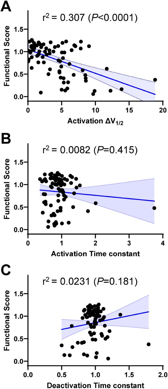
    Correlation of biophysical properties with functional scores from a KCNE1 deep mutation scan. ( A ) Plot of differences in activation V½ (ΔV½ in mV) measured in cells co-expressing WT KCNQ1 with KCNE1 variants (homozygous state) compared with the Functional Score reported for each variant determined by a cell fitness assay. ( B ) Plot of differences in activation time constants measured in cells co-expressing WT KCNQ1 with KCNE1 variants (homozygous state) compared with the reported Functional Score. ( C ) Plot of differences in deactivation time constants measured in cells co-expressing WT KCNQ1 with KCNE1 variants (homozygous state) compared with the reported Functional Score.

    Journal: bioRxiv

    Article Title: Functional profiling of KCNE1 variants informs population carrier frequency of Jervell and Lange-Nielsen syndrome type 2

    doi: 10.1101/2025.03.28.646046

    Figure Lengend Snippet: Correlation of biophysical properties with functional scores from a KCNE1 deep mutation scan. ( A ) Plot of differences in activation V½ (ΔV½ in mV) measured in cells co-expressing WT KCNQ1 with KCNE1 variants (homozygous state) compared with the Functional Score reported for each variant determined by a cell fitness assay. ( B ) Plot of differences in activation time constants measured in cells co-expressing WT KCNQ1 with KCNE1 variants (homozygous state) compared with the reported Functional Score. ( C ) Plot of differences in deactivation time constants measured in cells co-expressing WT KCNQ1 with KCNE1 variants (homozygous state) compared with the reported Functional Score.

    Article Snippet: Full length cDNA encoding WT KCNQ1 (GenBank accession number AF000571 ) was engineered in the mammalian expression vector pIRES-CyOFP1 (pIRES-CyOFP1-KCNQ1, AddGene #173162).

    Techniques: Functional Assay, Mutagenesis, Activation Assay, Expressing, Variant Assay