human pad2 protein Search Results


90
ModiQuest recombinant human pad2
Human MDMs (A) and mouse RAW 264.7 (B) macrophages were stimulated with 10 ng/mL LPS in the presence of native or citrullinated LL-37 at the indicated concentrations (0.1–10 μg/mL). Citrullinated LL-37 was obtained by treatment of the native peptide with human <t>PAD2</t> or PAD4 at 23.3 U/mg peptide). The level of TNF-α (A) and NO (B) in the culture supernatants was determined using ELISA or the Griess assay at 6 or 20 h post-stimulation, respectively. Since neither the LL-37 nor the PAD enzymes alone induced the release of NO or TNF-α, for the sake of clarity these controls are not shown in the figure. Data represent the mean ± SD of three independent experiments. ns, not significant; *, p<0.05; **, p<0.01; ***, p<0.001.
Recombinant Human Pad2, supplied by ModiQuest, used in various techniques. Bioz Stars score: 90/100, based on 1 PubMed citations. ZERO BIAS - scores, article reviews, protocol conditions and more
https://www.bioz.com/product/human+pad2+protein/recombinant+human+pad2/pmc04036085-68-0-8
Average 90 stars, based on 1 article reviews
recombinant human pad2 - by Bioz Stars, 2026-09
90/100 stars
  Buy from Supplier

94
Proteintech anti padi2
Human MDMs (A) and mouse RAW 264.7 (B) macrophages were stimulated with 10 ng/mL LPS in the presence of native or citrullinated LL-37 at the indicated concentrations (0.1–10 μg/mL). Citrullinated LL-37 was obtained by treatment of the native peptide with human <t>PAD2</t> or PAD4 at 23.3 U/mg peptide). The level of TNF-α (A) and NO (B) in the culture supernatants was determined using ELISA or the Griess assay at 6 or 20 h post-stimulation, respectively. Since neither the LL-37 nor the PAD enzymes alone induced the release of NO or TNF-α, for the sake of clarity these controls are not shown in the figure. Data represent the mean ± SD of three independent experiments. ns, not significant; *, p<0.05; **, p<0.01; ***, p<0.001.
Anti Padi2, supplied by Proteintech, used in various techniques. Bioz Stars score: 94/100, based on 1 PubMed citations. ZERO BIAS - scores, article reviews, protocol conditions and more
https://www.bioz.com/product/human+pad2+protein/PADI2+Antibody/pmc04772006-113-9-11
Average 94 stars, based on 1 article reviews
anti padi2 - by Bioz Stars, 2026-09
94/100 stars
  Buy from Supplier

95
Proteintech pad2
a, The enzymatic reaction mediated by PADs converts arginine to citrulline. b, c, Western blot images of <t>PAD2</t> and PAD4-mediated citrullination of H3 (b) and TDP-43 (c). In (c) arrows indicate the observed shift in TDP-43 molecular weight. d, Coomassie staining of PAD2 (bands 1, 4), PAD4 (bands 2, 6), unmodified TDP-43 (band 3), PAD2-mediated citR TDP-43 (band 5), and PAD4-mediated citR TDP-43 (band 7). e, Bar diagrams showing the MW shift (kDa) of TDP-43 protein following citrullination. f, Schematic representation of arginine epitopes positioned in the human TDP-43 protein sequence. Position of the 11 citrullinated arginine epitopes are indicated in red. g, Six out of eleven arginine epitopes susceptible to citrullination laid within common RX(X)R (red box) or RXG/RGGG (green box) motifs in TDP-43 sequence. h, MS/MS spectrum showing b- and y-ion coverage of modified citR83 peptide and extracted ion chromatograms (XICs) showed abundance and retention time of unmodified R83 vs. citR83 with PAD2 and PAD4 treatment (intact peptide monoisotopic m/z (+2) 719.8457, (+3) 480.2329). i, Retention time peaks for TDP-43 peptides surrounding the unmodified or modified R83 epitope and base peak m/z undergoing methionine (Met85) oxidation, citrullination or both showed reliable time separation.
Pad2, supplied by Proteintech, used in various techniques. Bioz Stars score: 95/100, based on 1 PubMed citations. ZERO BIAS - scores, article reviews, protocol conditions and more
https://www.bioz.com/product/human+pad2+protein/PADI4+Polyclonal+antibody/bio_rxiv__2025__02__28__639952-152-0-8
Average 95 stars, based on 1 article reviews
pad2 - by Bioz Stars, 2026-09
95/100 stars
  Buy from Supplier

90
Novus Biologicals pad2
a, The enzymatic reaction mediated by PADs converts arginine to citrulline. b, c, Western blot images of <t>PAD2</t> and PAD4-mediated citrullination of H3 (b) and TDP-43 (c). In (c) arrows indicate the observed shift in TDP-43 molecular weight. d, Coomassie staining of PAD2 (bands 1, 4), PAD4 (bands 2, 6), unmodified TDP-43 (band 3), PAD2-mediated citR TDP-43 (band 5), and PAD4-mediated citR TDP-43 (band 7). e, Bar diagrams showing the MW shift (kDa) of TDP-43 protein following citrullination. f, Schematic representation of arginine epitopes positioned in the human TDP-43 protein sequence. Position of the 11 citrullinated arginine epitopes are indicated in red. g, Six out of eleven arginine epitopes susceptible to citrullination laid within common RX(X)R (red box) or RXG/RGGG (green box) motifs in TDP-43 sequence. h, MS/MS spectrum showing b- and y-ion coverage of modified citR83 peptide and extracted ion chromatograms (XICs) showed abundance and retention time of unmodified R83 vs. citR83 with PAD2 and PAD4 treatment (intact peptide monoisotopic m/z (+2) 719.8457, (+3) 480.2329). i, Retention time peaks for TDP-43 peptides surrounding the unmodified or modified R83 epitope and base peak m/z undergoing methionine (Met85) oxidation, citrullination or both showed reliable time separation.
Pad2, supplied by Novus Biologicals, used in various techniques. Bioz Stars score: 90/100, based on 1 PubMed citations. ZERO BIAS - scores, article reviews, protocol conditions and more
https://www.bioz.com/product/human+pad2+protein/PADI2+Antibody+(4D4)/pmc03119113-170-27-34
Average 90 stars, based on 1 article reviews
pad2 - by Bioz Stars, 2026-09
90/100 stars
  Buy from Supplier

90
ProteoGenix murine pad2 pad4
IgG responses to PADs in C3H mice. IgG responses to murine <t>PAD2</t> or human PAD2 or human PAD4 were analyzed by ELISAs. Plates were coated with PADs and blocked with BSA. Sera from primed mice were obtained at 15, 35, 55, and 65 d postimmunization and were diluted at 1/40. ( A ) For PBS-immunized mice, 15 sera were tested for murine PAD2, 8 sera were tested for human PAD2, and 9 sera were tested for human PAD4. ( B ) For PAD-immunized mice, each serum was tested against the same PAD used for each immunization. After washing, peroxidase-conjugated antimurine IgG was added. The OD was read at 405 nm. The background OD was obtained by adding each serum to a well without PAD (negative). Positive sera were defined as an OD value higher than twice the background OD.
Murine Pad2 Pad4, supplied by ProteoGenix, used in various techniques. Bioz Stars score: 90/100, based on 1 PubMed citations. ZERO BIAS - scores, article reviews, protocol conditions and more
https://www.bioz.com/product/human+pad2+protein/murine+pad2+pad4/pmc05703315-112-0-13
Average 90 stars, based on 1 article reviews
murine pad2 pad4 - by Bioz Stars, 2026-09
90/100 stars
  Buy from Supplier

93
Cell Signaling Technology Inc pad2
a Immunofluorescence analysis showing efficient uptake of Alexa Fluor 488-labeled CitH3 (5 µg/mL) by wild-type (WT) BMDMs but not by Tlr2⁻/⁻ BMDMs. Cellular intensity of Alexa-488 was quantified ( n = 5). Statistical analysis was performed using two-sided, Unpaired t test with Welch’s correction. b Cytokine profiling of pro- and anti-inflammatory mediators (IL-6, IL-1β, IL-10, TNFα, IFN-α, IFN-β) in supernatants of WT and Tlr2⁻/⁻ BMDMs treated with H3 or CitH3 peptides (15 µg/mL) for 16 h ( n = per group). LPS (200 ng/mL) served as positive control. Statistical analysis was performed using two-way ANOVA with Sidak’s multiple comparisons test. c Immunocytochemistry demonstrating nuclear translocation of <t>PAD2</t> in BMDMs after 1 h exposure to CitH3 peptide (15 µg/mL). LPS (200 ng/mL) served as a positive control. PAD2 was stained with Alexa Fluor 488 (green), and nuclei were counterstained with DAPI (blue). Quantification of PAD2 nuclear localization was analyzed by one-way ANOVA followed by Tukey’s multiple comparisons test ( n = 3 per group). d Subcellular fractionation analysis confirming significant nuclear localization and citrullination of PAD2 in WT BMDMs following treatment with H3, CitH3, or LPS. Quantification was analyzed using one-way ANOVA followed by Tukey’s multiple comparisons test ( n = per group). e In vitro citrullination assay showing MPB-tagged PAD2-mediated citrullination of H3 in a Ca²⁺-dependent manner. EGTA-mediated Ca²⁺ chelation eliminated CitH3 production. Similar results from three independent replicates. f In vitro citrullination assay showing PAD2 citrullination. PAD2 auto-citrullination was confirmed as His-tagged recombinant PAD2 co-incubated with MBP-tagged PAD2 resulted in PAD2 citrullination. PAD4 also promotes PAD2 citrullination. Quantification was performed by Image J, citrullination signals were normalized to corresponding input signals and further normalized to the matching EGTA-treated group in each replicate experiments. Statistical analysis was performed using two-sided t-test.Data are presented as mean ± SD. Statistical significance in all panels was determined as follows: * p < 0.05, ** p < 0.01, *** p < 0.001, **** p < 0.0001. Source data are provided as a Source Data file.
Pad2, supplied by Cell Signaling Technology Inc, used in various techniques. Bioz Stars score: 93/100, based on 1 PubMed citations. ZERO BIAS - scores, article reviews, protocol conditions and more
https://www.bioz.com/product/human+pad2+protein/Phospho-MERIT40+(Ser29)+Antibody/pmc12343807-422-7-12
Average 93 stars, based on 1 article reviews
pad2 - by Bioz Stars, 2026-09
93/100 stars
  Buy from Supplier

86
Merck & Co human fibrinogen
a Immunofluorescence analysis showing efficient uptake of Alexa Fluor 488-labeled CitH3 (5 µg/mL) by wild-type (WT) BMDMs but not by Tlr2⁻/⁻ BMDMs. Cellular intensity of Alexa-488 was quantified ( n = 5). Statistical analysis was performed using two-sided, Unpaired t test with Welch’s correction. b Cytokine profiling of pro- and anti-inflammatory mediators (IL-6, IL-1β, IL-10, TNFα, IFN-α, IFN-β) in supernatants of WT and Tlr2⁻/⁻ BMDMs treated with H3 or CitH3 peptides (15 µg/mL) for 16 h ( n = per group). LPS (200 ng/mL) served as positive control. Statistical analysis was performed using two-way ANOVA with Sidak’s multiple comparisons test. c Immunocytochemistry demonstrating nuclear translocation of <t>PAD2</t> in BMDMs after 1 h exposure to CitH3 peptide (15 µg/mL). LPS (200 ng/mL) served as a positive control. PAD2 was stained with Alexa Fluor 488 (green), and nuclei were counterstained with DAPI (blue). Quantification of PAD2 nuclear localization was analyzed by one-way ANOVA followed by Tukey’s multiple comparisons test ( n = 3 per group). d Subcellular fractionation analysis confirming significant nuclear localization and citrullination of PAD2 in WT BMDMs following treatment with H3, CitH3, or LPS. Quantification was analyzed using one-way ANOVA followed by Tukey’s multiple comparisons test ( n = per group). e In vitro citrullination assay showing MPB-tagged PAD2-mediated citrullination of H3 in a Ca²⁺-dependent manner. EGTA-mediated Ca²⁺ chelation eliminated CitH3 production. Similar results from three independent replicates. f In vitro citrullination assay showing PAD2 citrullination. PAD2 auto-citrullination was confirmed as His-tagged recombinant PAD2 co-incubated with MBP-tagged PAD2 resulted in PAD2 citrullination. PAD4 also promotes PAD2 citrullination. Quantification was performed by Image J, citrullination signals were normalized to corresponding input signals and further normalized to the matching EGTA-treated group in each replicate experiments. Statistical analysis was performed using two-sided t-test.Data are presented as mean ± SD. Statistical significance in all panels was determined as follows: * p < 0.05, ** p < 0.01, *** p < 0.001, **** p < 0.0001. Source data are provided as a Source Data file.
Human Fibrinogen, supplied by Merck & Co, used in various techniques. Bioz Stars score: 86/100, based on 1 PubMed citations. ZERO BIAS - scores, article reviews, protocol conditions and more
https://www.bioz.com/product/human+pad2+protein/fibrinogen+human/bio_rxiv__64898__2026__02__12__705563-99-0-2
Average 86 stars, based on 1 article reviews
human fibrinogen - by Bioz Stars, 2026-09
86/100 stars
  Buy from Supplier

ln-18  (ATCC)
96
ATCC ln-18
a Immunofluorescence analysis showing efficient uptake of Alexa Fluor 488-labeled CitH3 (5 µg/mL) by wild-type (WT) BMDMs but not by Tlr2⁻/⁻ BMDMs. Cellular intensity of Alexa-488 was quantified ( n = 5). Statistical analysis was performed using two-sided, Unpaired t test with Welch’s correction. b Cytokine profiling of pro- and anti-inflammatory mediators (IL-6, IL-1β, IL-10, TNFα, IFN-α, IFN-β) in supernatants of WT and Tlr2⁻/⁻ BMDMs treated with H3 or CitH3 peptides (15 µg/mL) for 16 h ( n = per group). LPS (200 ng/mL) served as positive control. Statistical analysis was performed using two-way ANOVA with Sidak’s multiple comparisons test. c Immunocytochemistry demonstrating nuclear translocation of <t>PAD2</t> in BMDMs after 1 h exposure to CitH3 peptide (15 µg/mL). LPS (200 ng/mL) served as a positive control. PAD2 was stained with Alexa Fluor 488 (green), and nuclei were counterstained with DAPI (blue). Quantification of PAD2 nuclear localization was analyzed by one-way ANOVA followed by Tukey’s multiple comparisons test ( n = 3 per group). d Subcellular fractionation analysis confirming significant nuclear localization and citrullination of PAD2 in WT BMDMs following treatment with H3, CitH3, or LPS. Quantification was analyzed using one-way ANOVA followed by Tukey’s multiple comparisons test ( n = per group). e In vitro citrullination assay showing MPB-tagged PAD2-mediated citrullination of H3 in a Ca²⁺-dependent manner. EGTA-mediated Ca²⁺ chelation eliminated CitH3 production. Similar results from three independent replicates. f In vitro citrullination assay showing PAD2 citrullination. PAD2 auto-citrullination was confirmed as His-tagged recombinant PAD2 co-incubated with MBP-tagged PAD2 resulted in PAD2 citrullination. PAD4 also promotes PAD2 citrullination. Quantification was performed by Image J, citrullination signals were normalized to corresponding input signals and further normalized to the matching EGTA-treated group in each replicate experiments. Statistical analysis was performed using two-sided t-test.Data are presented as mean ± SD. Statistical significance in all panels was determined as follows: * p < 0.05, ** p < 0.01, *** p < 0.001, **** p < 0.0001. Source data are provided as a Source Data file.
Ln 18, supplied by ATCC, used in various techniques. Bioz Stars score: 96/100, based on 1 PubMed citations. ZERO BIAS - scores, article reviews, protocol conditions and more
https://www.bioz.com/product/human+pad2+protein/LN-18/custom%40crl-2610%4032105816
Average 96 stars, based on 1 article reviews
ln-18 - by Bioz Stars, 2026-09
96/100 stars
  Buy from Supplier

90
Talecris Biotherapeutics human serum albumin hsa
a Immunofluorescence analysis showing efficient uptake of Alexa Fluor 488-labeled CitH3 (5 µg/mL) by wild-type (WT) BMDMs but not by Tlr2⁻/⁻ BMDMs. Cellular intensity of Alexa-488 was quantified ( n = 5). Statistical analysis was performed using two-sided, Unpaired t test with Welch’s correction. b Cytokine profiling of pro- and anti-inflammatory mediators (IL-6, IL-1β, IL-10, TNFα, IFN-α, IFN-β) in supernatants of WT and Tlr2⁻/⁻ BMDMs treated with H3 or CitH3 peptides (15 µg/mL) for 16 h ( n = per group). LPS (200 ng/mL) served as positive control. Statistical analysis was performed using two-way ANOVA with Sidak’s multiple comparisons test. c Immunocytochemistry demonstrating nuclear translocation of <t>PAD2</t> in BMDMs after 1 h exposure to CitH3 peptide (15 µg/mL). LPS (200 ng/mL) served as a positive control. PAD2 was stained with Alexa Fluor 488 (green), and nuclei were counterstained with DAPI (blue). Quantification of PAD2 nuclear localization was analyzed by one-way ANOVA followed by Tukey’s multiple comparisons test ( n = 3 per group). d Subcellular fractionation analysis confirming significant nuclear localization and citrullination of PAD2 in WT BMDMs following treatment with H3, CitH3, or LPS. Quantification was analyzed using one-way ANOVA followed by Tukey’s multiple comparisons test ( n = per group). e In vitro citrullination assay showing MPB-tagged PAD2-mediated citrullination of H3 in a Ca²⁺-dependent manner. EGTA-mediated Ca²⁺ chelation eliminated CitH3 production. Similar results from three independent replicates. f In vitro citrullination assay showing PAD2 citrullination. PAD2 auto-citrullination was confirmed as His-tagged recombinant PAD2 co-incubated with MBP-tagged PAD2 resulted in PAD2 citrullination. PAD4 also promotes PAD2 citrullination. Quantification was performed by Image J, citrullination signals were normalized to corresponding input signals and further normalized to the matching EGTA-treated group in each replicate experiments. Statistical analysis was performed using two-sided t-test.Data are presented as mean ± SD. Statistical significance in all panels was determined as follows: * p < 0.05, ** p < 0.01, *** p < 0.001, **** p < 0.0001. Source data are provided as a Source Data file.
Human Serum Albumin Hsa, supplied by Talecris Biotherapeutics, used in various techniques. Bioz Stars score: 90/100, based on 1 PubMed citations. ZERO BIAS - scores, article reviews, protocol conditions and more
https://www.bioz.com/product/human+pad2+protein/human+serum+albumin/pm32224441-46-6-11
Average 90 stars, based on 1 article reviews
human serum albumin hsa - by Bioz Stars, 2026-09
90/100 stars
  Buy from Supplier

90
ModiQuest recombinant human pad4
a Immunofluorescence analysis showing efficient uptake of Alexa Fluor 488-labeled CitH3 (5 µg/mL) by wild-type (WT) BMDMs but not by Tlr2⁻/⁻ BMDMs. Cellular intensity of Alexa-488 was quantified ( n = 5). Statistical analysis was performed using two-sided, Unpaired t test with Welch’s correction. b Cytokine profiling of pro- and anti-inflammatory mediators (IL-6, IL-1β, IL-10, TNFα, IFN-α, IFN-β) in supernatants of WT and Tlr2⁻/⁻ BMDMs treated with H3 or CitH3 peptides (15 µg/mL) for 16 h ( n = per group). LPS (200 ng/mL) served as positive control. Statistical analysis was performed using two-way ANOVA with Sidak’s multiple comparisons test. c Immunocytochemistry demonstrating nuclear translocation of <t>PAD2</t> in BMDMs after 1 h exposure to CitH3 peptide (15 µg/mL). LPS (200 ng/mL) served as a positive control. PAD2 was stained with Alexa Fluor 488 (green), and nuclei were counterstained with DAPI (blue). Quantification of PAD2 nuclear localization was analyzed by one-way ANOVA followed by Tukey’s multiple comparisons test ( n = 3 per group). d Subcellular fractionation analysis confirming significant nuclear localization and citrullination of PAD2 in WT BMDMs following treatment with H3, CitH3, or LPS. Quantification was analyzed using one-way ANOVA followed by Tukey’s multiple comparisons test ( n = per group). e In vitro citrullination assay showing MPB-tagged PAD2-mediated citrullination of H3 in a Ca²⁺-dependent manner. EGTA-mediated Ca²⁺ chelation eliminated CitH3 production. Similar results from three independent replicates. f In vitro citrullination assay showing PAD2 citrullination. PAD2 auto-citrullination was confirmed as His-tagged recombinant PAD2 co-incubated with MBP-tagged PAD2 resulted in PAD2 citrullination. PAD4 also promotes PAD2 citrullination. Quantification was performed by Image J, citrullination signals were normalized to corresponding input signals and further normalized to the matching EGTA-treated group in each replicate experiments. Statistical analysis was performed using two-sided t-test.Data are presented as mean ± SD. Statistical significance in all panels was determined as follows: * p < 0.05, ** p < 0.01, *** p < 0.001, **** p < 0.0001. Source data are provided as a Source Data file.
Recombinant Human Pad4, supplied by ModiQuest, used in various techniques. Bioz Stars score: 90/100, based on 1 PubMed citations. ZERO BIAS - scores, article reviews, protocol conditions and more
https://www.bioz.com/product/human+pad2+protein/peptidylarginine+deiminase+4/pmc05036245__annrheumdis___2015___208495___s1-5-0-23
Average 90 stars, based on 1 article reviews
recombinant human pad4 - by Bioz Stars, 2026-09
90/100 stars
  Buy from Supplier

93
Cell Signaling Technology Inc anti pad2
a Immunofluorescence analysis showing efficient uptake of Alexa Fluor 488-labeled CitH3 (5 µg/mL) by wild-type (WT) BMDMs but not by Tlr2⁻/⁻ BMDMs. Cellular intensity of Alexa-488 was quantified ( n = 5). Statistical analysis was performed using two-sided, Unpaired t test with Welch’s correction. b Cytokine profiling of pro- and anti-inflammatory mediators (IL-6, IL-1β, IL-10, TNFα, IFN-α, IFN-β) in supernatants of WT and Tlr2⁻/⁻ BMDMs treated with H3 or CitH3 peptides (15 µg/mL) for 16 h ( n = per group). LPS (200 ng/mL) served as positive control. Statistical analysis was performed using two-way ANOVA with Sidak’s multiple comparisons test. c Immunocytochemistry demonstrating nuclear translocation of <t>PAD2</t> in BMDMs after 1 h exposure to CitH3 peptide (15 µg/mL). LPS (200 ng/mL) served as a positive control. PAD2 was stained with Alexa Fluor 488 (green), and nuclei were counterstained with DAPI (blue). Quantification of PAD2 nuclear localization was analyzed by one-way ANOVA followed by Tukey’s multiple comparisons test ( n = 3 per group). d Subcellular fractionation analysis confirming significant nuclear localization and citrullination of PAD2 in WT BMDMs following treatment with H3, CitH3, or LPS. Quantification was analyzed using one-way ANOVA followed by Tukey’s multiple comparisons test ( n = per group). e In vitro citrullination assay showing MPB-tagged PAD2-mediated citrullination of H3 in a Ca²⁺-dependent manner. EGTA-mediated Ca²⁺ chelation eliminated CitH3 production. Similar results from three independent replicates. f In vitro citrullination assay showing PAD2 citrullination. PAD2 auto-citrullination was confirmed as His-tagged recombinant PAD2 co-incubated with MBP-tagged PAD2 resulted in PAD2 citrullination. PAD4 also promotes PAD2 citrullination. Quantification was performed by Image J, citrullination signals were normalized to corresponding input signals and further normalized to the matching EGTA-treated group in each replicate experiments. Statistical analysis was performed using two-sided t-test.Data are presented as mean ± SD. Statistical significance in all panels was determined as follows: * p < 0.05, ** p < 0.01, *** p < 0.001, **** p < 0.0001. Source data are provided as a Source Data file.
Anti Pad2, supplied by Cell Signaling Technology Inc, used in various techniques. Bioz Stars score: 93/100, based on 1 PubMed citations. ZERO BIAS - scores, article reviews, protocol conditions and more
https://www.bioz.com/product/human+pad2+protein/PADI2+Rabbit+mAb/pmc10719474-63-33-35
Average 93 stars, based on 1 article reviews
anti pad2 - by Bioz Stars, 2026-09
93/100 stars
  Buy from Supplier

90
Merck KGaA human fibrinogen
Proliferation of T cells in response to human PAD 4, native <t>fibrinogen</t> ( FB ), citrullinated fibrinogen (cit FB), and phytohemagglutinin ( PHA ) in patients with RA , patients with PsA, and healthy controls. Proliferative response was evaluated by bromodeoxyuridine incorporation (n = 4 replicates per protein). The OD ratio was the ratio of the OD for a well with cells and protein to the OD for a well with cells but without protein. Positivity was defined as an OD ratio of >2 (dotted lines). Symbols represent individual subjects; red lines show the mean OD ratio. See Figure for other definitions.
Human Fibrinogen, supplied by Merck KGaA, used in various techniques. Bioz Stars score: 90/100, based on 1 PubMed citations. ZERO BIAS - scores, article reviews, protocol conditions and more
https://www.bioz.com/product/human+pad2+protein/human+fibrinogen/pmc07317357-44-0-2
Average 90 stars, based on 1 article reviews
human fibrinogen - by Bioz Stars, 2026-09
90/100 stars
  Buy from Supplier

Image Search Results


Human MDMs (A) and mouse RAW 264.7 (B) macrophages were stimulated with 10 ng/mL LPS in the presence of native or citrullinated LL-37 at the indicated concentrations (0.1–10 μg/mL). Citrullinated LL-37 was obtained by treatment of the native peptide with human PAD2 or PAD4 at 23.3 U/mg peptide). The level of TNF-α (A) and NO (B) in the culture supernatants was determined using ELISA or the Griess assay at 6 or 20 h post-stimulation, respectively. Since neither the LL-37 nor the PAD enzymes alone induced the release of NO or TNF-α, for the sake of clarity these controls are not shown in the figure. Data represent the mean ± SD of three independent experiments. ns, not significant; *, p<0.05; **, p<0.01; ***, p<0.001.

Journal: Journal of immunology (Baltimore, Md. : 1950)

Article Title: Citrullination alters immunomodulatory function of LL-37 essential for prevention of endotoxin-induced sepsis

doi: 10.4049/jimmunol.1303062

Figure Lengend Snippet: Human MDMs (A) and mouse RAW 264.7 (B) macrophages were stimulated with 10 ng/mL LPS in the presence of native or citrullinated LL-37 at the indicated concentrations (0.1–10 μg/mL). Citrullinated LL-37 was obtained by treatment of the native peptide with human PAD2 or PAD4 at 23.3 U/mg peptide). The level of TNF-α (A) and NO (B) in the culture supernatants was determined using ELISA or the Griess assay at 6 or 20 h post-stimulation, respectively. Since neither the LL-37 nor the PAD enzymes alone induced the release of NO or TNF-α, for the sake of clarity these controls are not shown in the figure. Data represent the mean ± SD of three independent experiments. ns, not significant; *, p<0.05; **, p<0.01; ***, p<0.001.

Article Snippet: Recombinant human PAD2 and PAD4 were obtained from Modiquest (The Netherlands).

Techniques: Enzyme-linked Immunosorbent Assay, Griess Assay

a, The enzymatic reaction mediated by PADs converts arginine to citrulline. b, c, Western blot images of PAD2 and PAD4-mediated citrullination of H3 (b) and TDP-43 (c). In (c) arrows indicate the observed shift in TDP-43 molecular weight. d, Coomassie staining of PAD2 (bands 1, 4), PAD4 (bands 2, 6), unmodified TDP-43 (band 3), PAD2-mediated citR TDP-43 (band 5), and PAD4-mediated citR TDP-43 (band 7). e, Bar diagrams showing the MW shift (kDa) of TDP-43 protein following citrullination. f, Schematic representation of arginine epitopes positioned in the human TDP-43 protein sequence. Position of the 11 citrullinated arginine epitopes are indicated in red. g, Six out of eleven arginine epitopes susceptible to citrullination laid within common RX(X)R (red box) or RXG/RGGG (green box) motifs in TDP-43 sequence. h, MS/MS spectrum showing b- and y-ion coverage of modified citR83 peptide and extracted ion chromatograms (XICs) showed abundance and retention time of unmodified R83 vs. citR83 with PAD2 and PAD4 treatment (intact peptide monoisotopic m/z (+2) 719.8457, (+3) 480.2329). i, Retention time peaks for TDP-43 peptides surrounding the unmodified or modified R83 epitope and base peak m/z undergoing methionine (Met85) oxidation, citrullination or both showed reliable time separation.

Journal: bioRxiv

Article Title: Citrullination of TDP-43 is a key post-translation modification associated with structural and functional changes and progressive pathology in TDP-43 mouse models and human proteinopathies

doi: 10.1101/2025.02.28.639952

Figure Lengend Snippet: a, The enzymatic reaction mediated by PADs converts arginine to citrulline. b, c, Western blot images of PAD2 and PAD4-mediated citrullination of H3 (b) and TDP-43 (c). In (c) arrows indicate the observed shift in TDP-43 molecular weight. d, Coomassie staining of PAD2 (bands 1, 4), PAD4 (bands 2, 6), unmodified TDP-43 (band 3), PAD2-mediated citR TDP-43 (band 5), and PAD4-mediated citR TDP-43 (band 7). e, Bar diagrams showing the MW shift (kDa) of TDP-43 protein following citrullination. f, Schematic representation of arginine epitopes positioned in the human TDP-43 protein sequence. Position of the 11 citrullinated arginine epitopes are indicated in red. g, Six out of eleven arginine epitopes susceptible to citrullination laid within common RX(X)R (red box) or RXG/RGGG (green box) motifs in TDP-43 sequence. h, MS/MS spectrum showing b- and y-ion coverage of modified citR83 peptide and extracted ion chromatograms (XICs) showed abundance and retention time of unmodified R83 vs. citR83 with PAD2 and PAD4 treatment (intact peptide monoisotopic m/z (+2) 719.8457, (+3) 480.2329). i, Retention time peaks for TDP-43 peptides surrounding the unmodified or modified R83 epitope and base peak m/z undergoing methionine (Met85) oxidation, citrullination or both showed reliable time separation.

Article Snippet: PAD2 and PAD4 primary antibodies were purchased from Proteintech, US (#12110-1-AP and 17373-1-AP) and diluted at 1:10000 vs. 1:1000, respectively.

Techniques: Western Blot, Molecular Weight, Staining, Sequencing, Tandem Mass Spectroscopy, Modification

Spectra showing b- and y-ion coverage of citrullinated peptides, as well as XICs retention times for unmodified and citrullinated peptides treated with PAD2 and PAD4, corresponding to a - c, citR165 (intact peptide monoisotopic m/z (+2) 665.8214), d - f, citR191(intact peptide monoisotopic m/z (+2) 672.8469), g - i, citR268/272 (intact peptide monoisotopic m/z (+2) 728.3560), j - l, citR293 (intact peptide monoisotopic m/z (+2) 638.29).

Journal: bioRxiv

Article Title: Citrullination of TDP-43 is a key post-translation modification associated with structural and functional changes and progressive pathology in TDP-43 mouse models and human proteinopathies

doi: 10.1101/2025.02.28.639952

Figure Lengend Snippet: Spectra showing b- and y-ion coverage of citrullinated peptides, as well as XICs retention times for unmodified and citrullinated peptides treated with PAD2 and PAD4, corresponding to a - c, citR165 (intact peptide monoisotopic m/z (+2) 665.8214), d - f, citR191(intact peptide monoisotopic m/z (+2) 672.8469), g - i, citR268/272 (intact peptide monoisotopic m/z (+2) 728.3560), j - l, citR293 (intact peptide monoisotopic m/z (+2) 638.29).

Article Snippet: PAD2 and PAD4 primary antibodies were purchased from Proteintech, US (#12110-1-AP and 17373-1-AP) and diluted at 1:10000 vs. 1:1000, respectively.

Techniques:

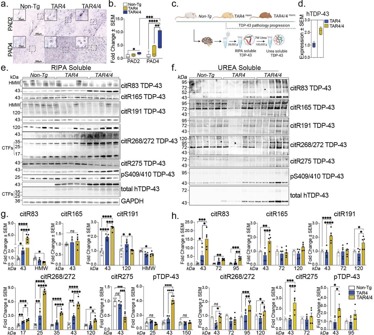
a, Immunohistochemical images of Non-Tg, TAR4 and TAR4/4 cortex labeled with PAD2 and PAD4 antibodies. b, Fold change of PAD2 and PAD4 (% area mean of the values ± SEM) normalized to the Non-Tg control. One-way or Two-way ANOVA, followed by Tukey’s post hoc multiple comparisons tests, n = 5, *p < 0.05, **p < 0.01, ***p < 0.001, ****p < 0.0001. c, Schematic overview of the TAR TDP-43 mouse model and the fractionation of cortical tissue into RIPA-soluble and (7 M) Urea-soluble fractions. d, The expression levels of the human TDP-43 in the TAR model. e, Cortical RIPA soluble and f, Urea soluble fraction analyzed by Western blotting and probed for citR TDP-43 antibody panel (citR83, 165, 191, 268/272, 275), pTDP-43 409/410 and total human TDP-43 protein. g, Quantification of citR TDP-43 43 kDa protein levels, proteolytic fragments (17, 25 & 35 kDa) and intermediate high molecular species (72, 98 & 120k Da) normalized to GAPDH in RIPA and h, Urea fraction normalized to the total protein loaded signal. Data represent the mean of the values ± SEM; One-way ANOVA, followed by Tukey’s or Šidák post hoc multiple comparisons tests, n = 5, *p < 0.05, **p < 0.01, ***p < 0.001, ****p < 0.0001.

Journal: bioRxiv

Article Title: Citrullination of TDP-43 is a key post-translation modification associated with structural and functional changes and progressive pathology in TDP-43 mouse models and human proteinopathies

doi: 10.1101/2025.02.28.639952

Figure Lengend Snippet: a, Immunohistochemical images of Non-Tg, TAR4 and TAR4/4 cortex labeled with PAD2 and PAD4 antibodies. b, Fold change of PAD2 and PAD4 (% area mean of the values ± SEM) normalized to the Non-Tg control. One-way or Two-way ANOVA, followed by Tukey’s post hoc multiple comparisons tests, n = 5, *p < 0.05, **p < 0.01, ***p < 0.001, ****p < 0.0001. c, Schematic overview of the TAR TDP-43 mouse model and the fractionation of cortical tissue into RIPA-soluble and (7 M) Urea-soluble fractions. d, The expression levels of the human TDP-43 in the TAR model. e, Cortical RIPA soluble and f, Urea soluble fraction analyzed by Western blotting and probed for citR TDP-43 antibody panel (citR83, 165, 191, 268/272, 275), pTDP-43 409/410 and total human TDP-43 protein. g, Quantification of citR TDP-43 43 kDa protein levels, proteolytic fragments (17, 25 & 35 kDa) and intermediate high molecular species (72, 98 & 120k Da) normalized to GAPDH in RIPA and h, Urea fraction normalized to the total protein loaded signal. Data represent the mean of the values ± SEM; One-way ANOVA, followed by Tukey’s or Šidák post hoc multiple comparisons tests, n = 5, *p < 0.05, **p < 0.01, ***p < 0.001, ****p < 0.0001.

Article Snippet: PAD2 and PAD4 primary antibodies were purchased from Proteintech, US (#12110-1-AP and 17373-1-AP) and diluted at 1:10000 vs. 1:1000, respectively.

Techniques: Immunohistochemical staining, Labeling, Control, Fractionation, Expressing, Western Blot

IgG responses to PADs in C3H mice. IgG responses to murine PAD2 or human PAD2 or human PAD4 were analyzed by ELISAs. Plates were coated with PADs and blocked with BSA. Sera from primed mice were obtained at 15, 35, 55, and 65 d postimmunization and were diluted at 1/40. ( A ) For PBS-immunized mice, 15 sera were tested for murine PAD2, 8 sera were tested for human PAD2, and 9 sera were tested for human PAD4. ( B ) For PAD-immunized mice, each serum was tested against the same PAD used for each immunization. After washing, peroxidase-conjugated antimurine IgG was added. The OD was read at 405 nm. The background OD was obtained by adding each serum to a well without PAD (negative). Positive sera were defined as an OD value higher than twice the background OD.

Journal: Proceedings of the National Academy of Sciences of the United States of America

Article Title: Peptidyl arginine deiminase immunization induces anticitrullinated protein antibodies in mice with particular MHC types

doi: 10.1073/pnas.1713112114

Figure Lengend Snippet: IgG responses to PADs in C3H mice. IgG responses to murine PAD2 or human PAD2 or human PAD4 were analyzed by ELISAs. Plates were coated with PADs and blocked with BSA. Sera from primed mice were obtained at 15, 35, 55, and 65 d postimmunization and were diluted at 1/40. ( A ) For PBS-immunized mice, 15 sera were tested for murine PAD2, 8 sera were tested for human PAD2, and 9 sera were tested for human PAD4. ( B ) For PAD-immunized mice, each serum was tested against the same PAD used for each immunization. After washing, peroxidase-conjugated antimurine IgG was added. The OD was read at 405 nm. The background OD was obtained by adding each serum to a well without PAD (negative). Positive sera were defined as an OD value higher than twice the background OD.

Article Snippet: Murine PAD2 and PAD4 and human PAD2 and PAD4 proteins were purchased from Proteogenix.

Techniques:

IgG responses to PADs in DBA/2 mice. Plates were coated with PADs and blocked with BSA. Sera from primed mice were obtained at 15, 35, 55, and 65 d postimmunization and were diluted at 1/40. ( A ) For PBS-immunized mice, nine sera were tested for murine PAD2 and human PAD4. ( B ) For PAD-immunized mice, each serum was tested against the same PAD used for each immunization. After washing, peroxidase-conjugated antimurine IgG was added. The OD was read at 405 nm. The background OD was obtained by adding each serum to a well without PAD (negative). Positive sera were defined as an OD value higher than twice the background OD.

Journal: Proceedings of the National Academy of Sciences of the United States of America

Article Title: Peptidyl arginine deiminase immunization induces anticitrullinated protein antibodies in mice with particular MHC types

doi: 10.1073/pnas.1713112114

Figure Lengend Snippet: IgG responses to PADs in DBA/2 mice. Plates were coated with PADs and blocked with BSA. Sera from primed mice were obtained at 15, 35, 55, and 65 d postimmunization and were diluted at 1/40. ( A ) For PBS-immunized mice, nine sera were tested for murine PAD2 and human PAD4. ( B ) For PAD-immunized mice, each serum was tested against the same PAD used for each immunization. After washing, peroxidase-conjugated antimurine IgG was added. The OD was read at 405 nm. The background OD was obtained by adding each serum to a well without PAD (negative). Positive sera were defined as an OD value higher than twice the background OD.

Article Snippet: Murine PAD2 and PAD4 and human PAD2 and PAD4 proteins were purchased from Proteogenix.

Techniques:

a Immunofluorescence analysis showing efficient uptake of Alexa Fluor 488-labeled CitH3 (5 µg/mL) by wild-type (WT) BMDMs but not by Tlr2⁻/⁻ BMDMs. Cellular intensity of Alexa-488 was quantified ( n = 5). Statistical analysis was performed using two-sided, Unpaired t test with Welch’s correction. b Cytokine profiling of pro- and anti-inflammatory mediators (IL-6, IL-1β, IL-10, TNFα, IFN-α, IFN-β) in supernatants of WT and Tlr2⁻/⁻ BMDMs treated with H3 or CitH3 peptides (15 µg/mL) for 16 h ( n = per group). LPS (200 ng/mL) served as positive control. Statistical analysis was performed using two-way ANOVA with Sidak’s multiple comparisons test. c Immunocytochemistry demonstrating nuclear translocation of PAD2 in BMDMs after 1 h exposure to CitH3 peptide (15 µg/mL). LPS (200 ng/mL) served as a positive control. PAD2 was stained with Alexa Fluor 488 (green), and nuclei were counterstained with DAPI (blue). Quantification of PAD2 nuclear localization was analyzed by one-way ANOVA followed by Tukey’s multiple comparisons test ( n = 3 per group). d Subcellular fractionation analysis confirming significant nuclear localization and citrullination of PAD2 in WT BMDMs following treatment with H3, CitH3, or LPS. Quantification was analyzed using one-way ANOVA followed by Tukey’s multiple comparisons test ( n = per group). e In vitro citrullination assay showing MPB-tagged PAD2-mediated citrullination of H3 in a Ca²⁺-dependent manner. EGTA-mediated Ca²⁺ chelation eliminated CitH3 production. Similar results from three independent replicates. f In vitro citrullination assay showing PAD2 citrullination. PAD2 auto-citrullination was confirmed as His-tagged recombinant PAD2 co-incubated with MBP-tagged PAD2 resulted in PAD2 citrullination. PAD4 also promotes PAD2 citrullination. Quantification was performed by Image J, citrullination signals were normalized to corresponding input signals and further normalized to the matching EGTA-treated group in each replicate experiments. Statistical analysis was performed using two-sided t-test.Data are presented as mean ± SD. Statistical significance in all panels was determined as follows: * p < 0.05, ** p < 0.01, *** p < 0.001, **** p < 0.0001. Source data are provided as a Source Data file.

Journal: Nature Communications

Article Title: A citrullinated histone H3 monoclonal antibody for immune modulation in sepsis

doi: 10.1038/s41467-025-62788-6

Figure Lengend Snippet: a Immunofluorescence analysis showing efficient uptake of Alexa Fluor 488-labeled CitH3 (5 µg/mL) by wild-type (WT) BMDMs but not by Tlr2⁻/⁻ BMDMs. Cellular intensity of Alexa-488 was quantified ( n = 5). Statistical analysis was performed using two-sided, Unpaired t test with Welch’s correction. b Cytokine profiling of pro- and anti-inflammatory mediators (IL-6, IL-1β, IL-10, TNFα, IFN-α, IFN-β) in supernatants of WT and Tlr2⁻/⁻ BMDMs treated with H3 or CitH3 peptides (15 µg/mL) for 16 h ( n = per group). LPS (200 ng/mL) served as positive control. Statistical analysis was performed using two-way ANOVA with Sidak’s multiple comparisons test. c Immunocytochemistry demonstrating nuclear translocation of PAD2 in BMDMs after 1 h exposure to CitH3 peptide (15 µg/mL). LPS (200 ng/mL) served as a positive control. PAD2 was stained with Alexa Fluor 488 (green), and nuclei were counterstained with DAPI (blue). Quantification of PAD2 nuclear localization was analyzed by one-way ANOVA followed by Tukey’s multiple comparisons test ( n = 3 per group). d Subcellular fractionation analysis confirming significant nuclear localization and citrullination of PAD2 in WT BMDMs following treatment with H3, CitH3, or LPS. Quantification was analyzed using one-way ANOVA followed by Tukey’s multiple comparisons test ( n = per group). e In vitro citrullination assay showing MPB-tagged PAD2-mediated citrullination of H3 in a Ca²⁺-dependent manner. EGTA-mediated Ca²⁺ chelation eliminated CitH3 production. Similar results from three independent replicates. f In vitro citrullination assay showing PAD2 citrullination. PAD2 auto-citrullination was confirmed as His-tagged recombinant PAD2 co-incubated with MBP-tagged PAD2 resulted in PAD2 citrullination. PAD4 also promotes PAD2 citrullination. Quantification was performed by Image J, citrullination signals were normalized to corresponding input signals and further normalized to the matching EGTA-treated group in each replicate experiments. Statistical analysis was performed using two-sided t-test.Data are presented as mean ± SD. Statistical significance in all panels was determined as follows: * p < 0.05, ** p < 0.01, *** p < 0.001, **** p < 0.0001. Source data are provided as a Source Data file.

Article Snippet: The primary antibodies used in immunocytochemistry assays: PAD2 (12110-1-AP), PAD4 (17373-1-AP), Rab5 (CST, 3547), EEA1 (CST, 3288), MPO (Abcam ab208670)

Techniques: Immunofluorescence, Labeling, Positive Control, Immunocytochemistry, Translocation Assay, Staining, Fractionation, In Vitro, Recombinant, Incubation

a LDH release from supernatants of BMDMs treated with increasing doses of CitH3 or unmodified H3 peptides for 24 h at 37 °C, showing CitH3-induced cytotoxicity ( n = per group). b Cell viability, assessed using the CCK8 assay, demonstrated significant reduction following CitH3 peptide treatment compared to the H3 peptide ( n = 5 per group). c BMDMs treated with 50 µg/mL CitH3 peptide and increasing doses of hCitH3-mAb or human IgG. LDH release was measured to evaluate cytotoxicity ( n = per group). d , Viability of BMDMs treated as in ( c ) was measured using the CCK8 assay. hCitH3-mAb significantly preserved cell viability compared to control IgG ( n = per group). e Immunoblot analysis of CitH3 in BMDMs treated with 15 μg/mL CitH3 or H3 peptides. Cells were washed three times prior to collection. And given that the CitH3 peptide is only ~30 amino acids in length, the bands observed at ~17 kDa are interpreted as endogenous cellular CitH3 protein detected at various time points. Similar results from three independent replicates. f Western blot analysis of citrullinated proteins in BMDMs from WT, Pad2⁻/⁻, and Pad2/4⁻/⁻ mice following 1 h treatment with 15 μg/mL CitH3 peptide. CitH3-induced citrullination was PAD2-dependent. Similar results from four independent replicates. g Quantification of IL-6, IL-1β, TNFα, IFN-α, and IFN-β levels in the supernatants of BMDMs treated with 25 μg/mL CitH3 peptide for 24 h ( n = 6 per group). hCitH3-mAb effectively reduced CitH3-induced cytokine elevation in a dose-dependent manner, whereas control IgG had no effect. Data are presented as mean ± SD. Statistical analysis: One-way ANOVA followed by Dunnett’s multiple comparisons test was applied throughout. Comparisons were made to the ‘0’ group in ( a –d ), and the CitH3-treated group in ( g ), as indicated. Significance thresholds: * p < 0.05, ** p < 0.01, *** p < 0.001, **** p < 0.0001.Source data are provided as a Source Data file.

Journal: Nature Communications

Article Title: A citrullinated histone H3 monoclonal antibody for immune modulation in sepsis

doi: 10.1038/s41467-025-62788-6

Figure Lengend Snippet: a LDH release from supernatants of BMDMs treated with increasing doses of CitH3 or unmodified H3 peptides for 24 h at 37 °C, showing CitH3-induced cytotoxicity ( n = per group). b Cell viability, assessed using the CCK8 assay, demonstrated significant reduction following CitH3 peptide treatment compared to the H3 peptide ( n = 5 per group). c BMDMs treated with 50 µg/mL CitH3 peptide and increasing doses of hCitH3-mAb or human IgG. LDH release was measured to evaluate cytotoxicity ( n = per group). d , Viability of BMDMs treated as in ( c ) was measured using the CCK8 assay. hCitH3-mAb significantly preserved cell viability compared to control IgG ( n = per group). e Immunoblot analysis of CitH3 in BMDMs treated with 15 μg/mL CitH3 or H3 peptides. Cells were washed three times prior to collection. And given that the CitH3 peptide is only ~30 amino acids in length, the bands observed at ~17 kDa are interpreted as endogenous cellular CitH3 protein detected at various time points. Similar results from three independent replicates. f Western blot analysis of citrullinated proteins in BMDMs from WT, Pad2⁻/⁻, and Pad2/4⁻/⁻ mice following 1 h treatment with 15 μg/mL CitH3 peptide. CitH3-induced citrullination was PAD2-dependent. Similar results from four independent replicates. g Quantification of IL-6, IL-1β, TNFα, IFN-α, and IFN-β levels in the supernatants of BMDMs treated with 25 μg/mL CitH3 peptide for 24 h ( n = 6 per group). hCitH3-mAb effectively reduced CitH3-induced cytokine elevation in a dose-dependent manner, whereas control IgG had no effect. Data are presented as mean ± SD. Statistical analysis: One-way ANOVA followed by Dunnett’s multiple comparisons test was applied throughout. Comparisons were made to the ‘0’ group in ( a –d ), and the CitH3-treated group in ( g ), as indicated. Significance thresholds: * p < 0.05, ** p < 0.01, *** p < 0.001, **** p < 0.0001.Source data are provided as a Source Data file.

Article Snippet: The primary antibodies used in immunocytochemistry assays: PAD2 (12110-1-AP), PAD4 (17373-1-AP), Rab5 (CST, 3547), EEA1 (CST, 3288), MPO (Abcam ab208670)

Techniques: CCK-8 Assay, Control, Western Blot

Proliferation of T cells in response to human PAD 4, native fibrinogen ( FB ), citrullinated fibrinogen (cit FB), and phytohemagglutinin ( PHA ) in patients with RA , patients with PsA, and healthy controls. Proliferative response was evaluated by bromodeoxyuridine incorporation (n = 4 replicates per protein). The OD ratio was the ratio of the OD for a well with cells and protein to the OD for a well with cells but without protein. Positivity was defined as an OD ratio of >2 (dotted lines). Symbols represent individual subjects; red lines show the mean OD ratio. See Figure for other definitions.

Journal: Arthritis & Rheumatology (Hoboken, N.j.)

Article Title: Peptidylarginine Deiminase Autoimmunity and the Development of Anti–Citrullinated Protein Antibody in Rheumatoid Arthritis: The Hapten–Carrier Model

doi: 10.1002/art.41189

Figure Lengend Snippet: Proliferation of T cells in response to human PAD 4, native fibrinogen ( FB ), citrullinated fibrinogen (cit FB), and phytohemagglutinin ( PHA ) in patients with RA , patients with PsA, and healthy controls. Proliferative response was evaluated by bromodeoxyuridine incorporation (n = 4 replicates per protein). The OD ratio was the ratio of the OD for a well with cells and protein to the OD for a well with cells but without protein. Positivity was defined as an OD ratio of >2 (dotted lines). Symbols represent individual subjects; red lines show the mean OD ratio. See Figure for other definitions.

Article Snippet: Human fibrinogen (Merck Millipore) was incubated in a buffer containing 1 M Tris HCl (pH 7.4), 100 m M CaCl 2 , and 50 m M dithiothreitol at a concentration of 1 mg/ml with rabbit PAD2 protein (Sigma‐Aldrich).

Techniques: