c caspase 3 affinity Search Results


99
Cell Signaling Technology Inc c caspase 3
C Caspase 3, supplied by Cell Signaling Technology Inc, used in various techniques. Bioz Stars score: 99/100, based on 1 PubMed citations. ZERO BIAS - scores, article reviews, protocol conditions and more
https://www.bioz.com/product/c+caspase+3+affinity/Caspase-3+Antibody/ppr0762015-53-31-34
Average 99 stars, based on 1 article reviews
c caspase 3 - by Bioz Stars, 2026-10
99/100 stars
  Buy from Supplier

86
Affinity Biosciences c caspase3
COPH could decreases neuronal apoptosis at 7 d.p.i. (A) Immunofluorescence staining of NeuN (green), <t>C-caspase3</t> (red) and DAPI (blue) in different groups. Scale bar: 50 μm. (B) Western blotting showing the expression of Bax, Bcl-2 and C-caspase3 in different groups 7 d.p.i. (C) Statistical analysis of C-caspase3 fluorescence intensity in each group. (D–F) The quantitative analysis of Bax, Bcl-2 and C-caspase3 protein expression. ∗∗ represents P < 0.01 vs SCI group. ## represent COPs group vs COPH group respectively p < 0.01, (n = 3). (For interpretation of the references to colour in this figure legend, the reader is referred to the Web version of this article.)
C Caspase3, supplied by Affinity Biosciences, used in various techniques. Bioz Stars score: 86/100, based on 1 PubMed citations. ZERO BIAS - scores, article reviews, protocol conditions and more
https://www.bioz.com/product/c+caspase+3+affinity/3+anti+caspase+cleaved/pmc12857284-58-3-12
Average 86 stars, based on 1 article reviews
c caspase3 - by Bioz Stars, 2026-10
86/100 stars
  Buy from Supplier

90
Becton Dickinson anti-active caspase 3
COPH could decreases neuronal apoptosis at 7 d.p.i. (A) Immunofluorescence staining of NeuN (green), <t>C-caspase3</t> (red) and DAPI (blue) in different groups. Scale bar: 50 μm. (B) Western blotting showing the expression of Bax, Bcl-2 and C-caspase3 in different groups 7 d.p.i. (C) Statistical analysis of C-caspase3 fluorescence intensity in each group. (D–F) The quantitative analysis of Bax, Bcl-2 and C-caspase3 protein expression. ∗∗ represents P < 0.01 vs SCI group. ## represent COPs group vs COPH group respectively p < 0.01, (n = 3). (For interpretation of the references to colour in this figure legend, the reader is referred to the Web version of this article.)
Anti Active Caspase 3, supplied by Becton Dickinson, used in various techniques. Bioz Stars score: 90/100, based on 1 PubMed citations. ZERO BIAS - scores, article reviews, protocol conditions and more
https://www.bioz.com/product/c+caspase+3+affinity/anti+caspase+3/pm16818780-83-27-33
Average 90 stars, based on 1 article reviews
anti-active caspase 3 - by Bioz Stars, 2026-10
90/100 stars
  Buy from Supplier

97
Proteintech anti caspase 3
COPH could decreases neuronal apoptosis at 7 d.p.i. (A) Immunofluorescence staining of NeuN (green), <t>C-caspase3</t> (red) and DAPI (blue) in different groups. Scale bar: 50 μm. (B) Western blotting showing the expression of Bax, Bcl-2 and C-caspase3 in different groups 7 d.p.i. (C) Statistical analysis of C-caspase3 fluorescence intensity in each group. (D–F) The quantitative analysis of Bax, Bcl-2 and C-caspase3 protein expression. ∗∗ represents P < 0.01 vs SCI group. ## represent COPs group vs COPH group respectively p < 0.01, (n = 3). (For interpretation of the references to colour in this figure legend, the reader is referred to the Web version of this article.)
Anti Caspase 3, supplied by Proteintech, used in various techniques. Bioz Stars score: 97/100, based on 1 PubMed citations. ZERO BIAS - scores, article reviews, protocol conditions and more
https://www.bioz.com/product/c+caspase+3+affinity/Caspase+3%2Fp17%2Fp19+Antibody/pm36099983-127-20-50
Average 97 stars, based on 1 article reviews
anti caspase 3 - by Bioz Stars, 2026-10
97/100 stars
  Buy from Supplier

93
Cell Signaling Technology Inc rabbit polyclonal anti caspase 3
COPH could decreases neuronal apoptosis at 7 d.p.i. (A) Immunofluorescence staining of NeuN (green), <t>C-caspase3</t> (red) and DAPI (blue) in different groups. Scale bar: 50 μm. (B) Western blotting showing the expression of Bax, Bcl-2 and C-caspase3 in different groups 7 d.p.i. (C) Statistical analysis of C-caspase3 fluorescence intensity in each group. (D–F) The quantitative analysis of Bax, Bcl-2 and C-caspase3 protein expression. ∗∗ represents P < 0.01 vs SCI group. ## represent COPs group vs COPH group respectively p < 0.01, (n = 3). (For interpretation of the references to colour in this figure legend, the reader is referred to the Web version of this article.)
Rabbit Polyclonal Anti Caspase 3, supplied by Cell Signaling Technology Inc, used in various techniques. Bioz Stars score: 93/100, based on 1 PubMed citations. ZERO BIAS - scores, article reviews, protocol conditions and more
https://www.bioz.com/product/c+caspase+3+affinity/Epitope+Tag+Antibody+Sampler+Kit/pmc03946191-279-13-18
Average 93 stars, based on 1 article reviews
rabbit polyclonal anti caspase 3 - by Bioz Stars, 2026-10
93/100 stars
  Buy from Supplier

92
R&D Systems anti human mouse active caspase 3 antibody
COPH could decreases neuronal apoptosis at 7 d.p.i. (A) Immunofluorescence staining of NeuN (green), <t>C-caspase3</t> (red) and DAPI (blue) in different groups. Scale bar: 50 μm. (B) Western blotting showing the expression of Bax, Bcl-2 and C-caspase3 in different groups 7 d.p.i. (C) Statistical analysis of C-caspase3 fluorescence intensity in each group. (D–F) The quantitative analysis of Bax, Bcl-2 and C-caspase3 protein expression. ∗∗ represents P < 0.01 vs SCI group. ## represent COPs group vs COPH group respectively p < 0.01, (n = 3). (For interpretation of the references to colour in this figure legend, the reader is referred to the Web version of this article.)
Anti Human Mouse Active Caspase 3 Antibody, supplied by R&D Systems, used in various techniques. Bioz Stars score: 92/100, based on 1 PubMed citations. ZERO BIAS - scores, article reviews, protocol conditions and more
https://www.bioz.com/product/c+caspase+3+affinity/Human+Caspase-3+Antibody/pmc02743138-103-13-17
Average 92 stars, based on 1 article reviews
anti human mouse active caspase 3 antibody - by Bioz Stars, 2026-10
92/100 stars
  Buy from Supplier

96
Boster Bio antibody against c caspase 3
COPH could decreases neuronal apoptosis at 7 d.p.i. (A) Immunofluorescence staining of NeuN (green), <t>C-caspase3</t> (red) and DAPI (blue) in different groups. Scale bar: 50 μm. (B) Western blotting showing the expression of Bax, Bcl-2 and C-caspase3 in different groups 7 d.p.i. (C) Statistical analysis of C-caspase3 fluorescence intensity in each group. (D–F) The quantitative analysis of Bax, Bcl-2 and C-caspase3 protein expression. ∗∗ represents P < 0.01 vs SCI group. ## represent COPs group vs COPH group respectively p < 0.01, (n = 3). (For interpretation of the references to colour in this figure legend, the reader is referred to the Web version of this article.)
Antibody Against C Caspase 3, supplied by Boster Bio, used in various techniques. Bioz Stars score: 96/100, based on 1 PubMed citations. ZERO BIAS - scores, article reviews, protocol conditions and more
https://www.bioz.com/product/c+caspase+3+affinity/Anti-Caspase-3+CASP3+Antibody/pmc11299798-82-7-27
Average 96 stars, based on 1 article reviews
antibody against c caspase 3 - by Bioz Stars, 2026-10
96/100 stars
  Buy from Supplier

86
Wuhan Sanying Biotechnology c caspase3
Effects of HAP on cartilage autophagy and apoptosis in KOA model rats. Note: (A,C–F) Expression of LC3 I, LC3 II, Beclin1, <t>C-Caspase3</t> and Bax in cartilage tissue (GAPDH as internal control). (B,G,H) Expression of p62 and Bcl2 in cartilage (GAPDH as internal control). *** p < 0.001, ** p < 0.01, * p < 0.05.
C Caspase3, supplied by Wuhan Sanying Biotechnology, used in various techniques. Bioz Stars score: 86/100, based on 1 PubMed citations. ZERO BIAS - scores, article reviews, protocol conditions and more
https://www.bioz.com/product/c+caspase+3+affinity/3+anti+caspase/pmc13147163-122-23-27
Average 86 stars, based on 1 article reviews
c caspase3 - by Bioz Stars, 2026-10
86/100 stars
  Buy from Supplier

97
Cell Signaling Technology Inc tbst
Effects of HAP on cartilage autophagy and apoptosis in KOA model rats. Note: (A,C–F) Expression of LC3 I, LC3 II, Beclin1, <t>C-Caspase3</t> and Bax in cartilage tissue (GAPDH as internal control). (B,G,H) Expression of p62 and Bcl2 in cartilage (GAPDH as internal control). *** p < 0.001, ** p < 0.01, * p < 0.05.
Tbst, supplied by Cell Signaling Technology Inc, used in various techniques. Bioz Stars score: 97/100, based on 1 PubMed citations. ZERO BIAS - scores, article reviews, protocol conditions and more
https://www.bioz.com/product/c+caspase+3+affinity/Caspase-1+Antibody/pmc02722704-91-43-51
Average 97 stars, based on 1 article reviews
tbst - by Bioz Stars, 2026-10
97/100 stars
  Buy from Supplier

96
Proteintech 4◦c
Effects of HAP on cartilage autophagy and apoptosis in KOA model rats. Note: (A,C–F) Expression of LC3 I, LC3 II, Beclin1, <t>C-Caspase3</t> and Bax in cartilage tissue (GAPDH as internal control). (B,G,H) Expression of p62 and Bcl2 in cartilage (GAPDH as internal control). *** p < 0.001, ** p < 0.01, * p < 0.05.
4◦C, supplied by Proteintech, used in various techniques. Bioz Stars score: 96/100, based on 1 PubMed citations. ZERO BIAS - scores, article reviews, protocol conditions and more
https://www.bioz.com/product/c+caspase+3+affinity/Rac1+Antibody/pm39765117-79-22-25
Average 96 stars, based on 1 article reviews
4◦c - by Bioz Stars, 2026-10
96/100 stars
  Buy from Supplier

96
R&D Systems primary antibody anti caspase 3
Effects of HAP on cartilage autophagy and apoptosis in KOA model rats. Note: (A,C–F) Expression of LC3 I, LC3 II, Beclin1, <t>C-Caspase3</t> and Bax in cartilage tissue (GAPDH as internal control). (B,G,H) Expression of p62 and Bcl2 in cartilage (GAPDH as internal control). *** p < 0.001, ** p < 0.01, * p < 0.05.
Primary Antibody Anti Caspase 3, supplied by R&D Systems, used in various techniques. Bioz Stars score: 96/100, based on 1 PubMed citations. ZERO BIAS - scores, article reviews, protocol conditions and more
https://www.bioz.com/product/c+caspase+3+affinity/Human%2FMouse+Active+Caspase-3+Antibody/10__1128_slash_jvi__80__10__4801___4819__2006-150-44-55
Average 96 stars, based on 1 article reviews
primary antibody anti caspase 3 - by Bioz Stars, 2026-10
96/100 stars
  Buy from Supplier

96
Cell Signaling Technology Inc immunocytochemistry rabbit anti cleaved caspase 3
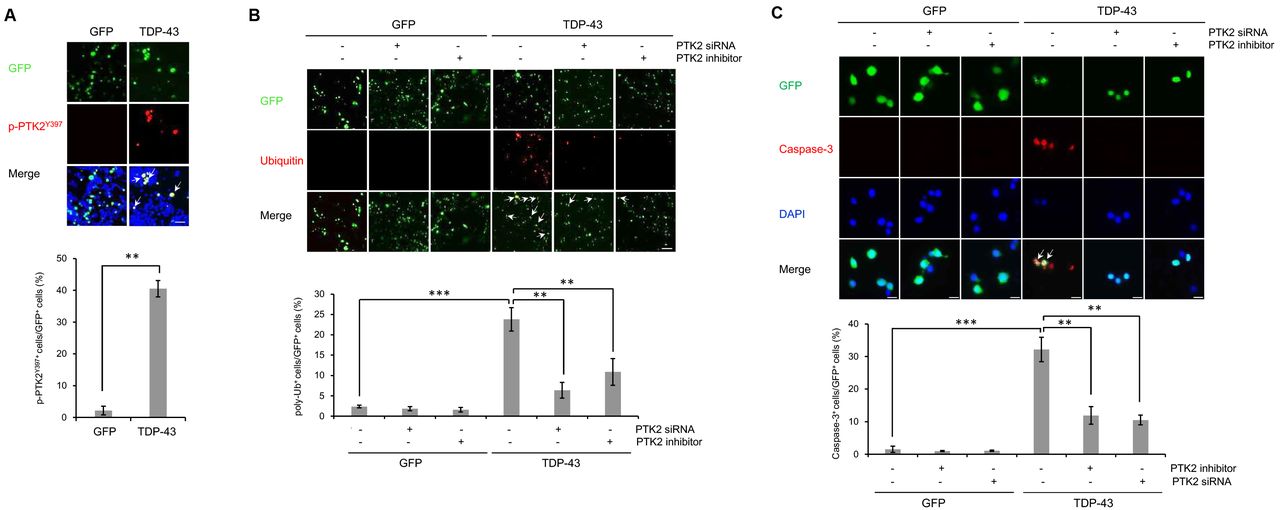
(A-C) N2a cells were transiently transfected with the control vector (pCMV6-AC-GFP) or TDP-43 expression construct (pCMV6-AC-TDP-43-GFP) for 3 days. Cells overexpressing GFP or TDP-43-GFP were treated with ptk2 -specific siRNA (20 nM) or a PTK2 inhibitor (5 μm) for 24 h. Immunocytochemistry was performed thereafter. (A) Immunocytochemistry was subsequently performed to detect PTK2 phosphorylation (p-PTK2 Y397 ; red). Quantification of the percentage of p-PTK2 Y397 -positive cells in GFP-positive cells ( lower ). Arrowheads indicate the co-localization of the p-PTK2 Y397 with TDP-43-GFP-positive cells. Data are presented as the means ± SD of 3 independent experiments. **p<0.005 (Student’s t -test). Scale bars, 200 μm. (B) Immunocytochemistry was subsequently performed to detect ubiquitin (red) or DAPI (nuclei; blue). Quantification of the percentage of poly-ubiquitin-positive staining in GFP-positive cells ( lower ). Arrowheads indicate the co-localization of the poly-ubiquitin with TDP-43-GFP-positive cells. Data are presented as the means ± SD of 3 independent experiments. **p<0.005, ***p<0.001 (two-way ANOVA with Bonferroni multiple comparison test). Scale bars, 20 μm. (C) Immunocytochemistry was subsequently performed to detect the expression of cleaved <t>caspase-3</t> (red) or DAPI (nuclei; blue). Quantification of the percentage of cleaved caspase-3-positive staining in GFP-positive cells (lower). Arrowheads indicate the co-localization of the caspase-3 with TDP-43-GFP-positive cells. Data are presented as the means ± SD of 3 independent experiments. **p<0.005, ***p<0.001 (two-way ANOVA with Bonferroni multiple comparison test). Scale bars, 200 μm.
Immunocytochemistry Rabbit Anti Cleaved Caspase 3, supplied by Cell Signaling Technology Inc, used in various techniques. Bioz Stars score: 96/100, based on 1 PubMed citations. ZERO BIAS - scores, article reviews, protocol conditions and more
https://www.bioz.com/product/c+caspase+3+affinity/Phospho-(Ser%2FThr)+Akt+Substrate+Antibody/bio_rxiv__355446-33-6-13
Average 96 stars, based on 1 article reviews
immunocytochemistry rabbit anti cleaved caspase 3 - by Bioz Stars, 2026-10
96/100 stars
  Buy from Supplier

Image Search Results


COPH could decreases neuronal apoptosis at 7 d.p.i. (A) Immunofluorescence staining of NeuN (green), C-caspase3 (red) and DAPI (blue) in different groups. Scale bar: 50 μm. (B) Western blotting showing the expression of Bax, Bcl-2 and C-caspase3 in different groups 7 d.p.i. (C) Statistical analysis of C-caspase3 fluorescence intensity in each group. (D–F) The quantitative analysis of Bax, Bcl-2 and C-caspase3 protein expression. ∗∗ represents P < 0.01 vs SCI group. ## represent COPs group vs COPH group respectively p < 0.01, (n = 3). (For interpretation of the references to colour in this figure legend, the reader is referred to the Web version of this article.)

Journal: Materials Today Bio

Article Title: A multifunctional and ROS response CO-gas delivery platform for spinal cord regeneration

doi: 10.1016/j.mtbio.2026.102760

Figure Lengend Snippet: COPH could decreases neuronal apoptosis at 7 d.p.i. (A) Immunofluorescence staining of NeuN (green), C-caspase3 (red) and DAPI (blue) in different groups. Scale bar: 50 μm. (B) Western blotting showing the expression of Bax, Bcl-2 and C-caspase3 in different groups 7 d.p.i. (C) Statistical analysis of C-caspase3 fluorescence intensity in each group. (D–F) The quantitative analysis of Bax, Bcl-2 and C-caspase3 protein expression. ∗∗ represents P < 0.01 vs SCI group. ## represent COPs group vs COPH group respectively p < 0.01, (n = 3). (For interpretation of the references to colour in this figure legend, the reader is referred to the Web version of this article.)

Article Snippet: Primary antibody to C-Caspase3, CD206, IL-1β, HO-1, SOD1, P65 was provided by Affinity Biosciences (USA).

Techniques: Immunofluorescence, Staining, Western Blot, Expressing, Fluorescence

Effects of HAP on cartilage autophagy and apoptosis in KOA model rats. Note: (A,C–F) Expression of LC3 I, LC3 II, Beclin1, C-Caspase3 and Bax in cartilage tissue (GAPDH as internal control). (B,G,H) Expression of p62 and Bcl2 in cartilage (GAPDH as internal control). *** p < 0.001, ** p < 0.01, * p < 0.05.

Journal: Frontiers in Bioengineering and Biotechnology

Article Title: Uncovering the mechanisms of homologous point acupuncture on knee osteoarthritis through an integrated study of metabolomics and proteomics

doi: 10.3389/fbioe.2026.1791109

Figure Lengend Snippet: Effects of HAP on cartilage autophagy and apoptosis in KOA model rats. Note: (A,C–F) Expression of LC3 I, LC3 II, Beclin1, C-Caspase3 and Bax in cartilage tissue (GAPDH as internal control). (B,G,H) Expression of p62 and Bcl2 in cartilage (GAPDH as internal control). *** p < 0.001, ** p < 0.01, * p < 0.05.

Article Snippet: All the primary antibodies were validated by either the manufacturer or other available publications, listed as follows: HIF1α (Boster, A00013-1), HK2 (Abclonal, A0994), c-caspase3 (Affinity, AF7022), Bax (Wuhan Sanying, 50599-2-Ig), LC3 (CST, 12741T), Beclin1 (Wuhan Sanying, 11306-1-AP), and GAPDH (Wuhan Sanying, 60004-1-Ig) from WUHAN HUAYAN Biotechnology CO., LTD. NLRP3 (Affinity, BF8029), ASC (Affinity, DF6304), caspase1 (Affinity, AF5418), p62 (Wuhan Sanying, 18420-1-AP), Bcl2 (Wuhan Sanying, 60178-1-Ig) from WUHAN Fabre Biotechnology CO., LTD. After washing 5 times for 5 min each with TBST (T-Pro), and a horseradish peroxidase (HRP)-conjugated secondary antibody (Boster, BA1051 and BA1054) was applied for 2 h at room temperature.

Techniques: Expressing, Control

(A-C) N2a cells were transiently transfected with the control vector (pCMV6-AC-GFP) or TDP-43 expression construct (pCMV6-AC-TDP-43-GFP) for 3 days. Cells overexpressing GFP or TDP-43-GFP were treated with ptk2 -specific siRNA (20 nM) or a PTK2 inhibitor (5 μm) for 24 h. Immunocytochemistry was performed thereafter. (A) Immunocytochemistry was subsequently performed to detect PTK2 phosphorylation (p-PTK2 Y397 ; red). Quantification of the percentage of p-PTK2 Y397 -positive cells in GFP-positive cells ( lower ). Arrowheads indicate the co-localization of the p-PTK2 Y397 with TDP-43-GFP-positive cells. Data are presented as the means ± SD of 3 independent experiments. **p<0.005 (Student’s t -test). Scale bars, 200 μm. (B) Immunocytochemistry was subsequently performed to detect ubiquitin (red) or DAPI (nuclei; blue). Quantification of the percentage of poly-ubiquitin-positive staining in GFP-positive cells ( lower ). Arrowheads indicate the co-localization of the poly-ubiquitin with TDP-43-GFP-positive cells. Data are presented as the means ± SD of 3 independent experiments. **p<0.005, ***p<0.001 (two-way ANOVA with Bonferroni multiple comparison test). Scale bars, 20 μm. (C) Immunocytochemistry was subsequently performed to detect the expression of cleaved caspase-3 (red) or DAPI (nuclei; blue). Quantification of the percentage of cleaved caspase-3-positive staining in GFP-positive cells (lower). Arrowheads indicate the co-localization of the caspase-3 with TDP-43-GFP-positive cells. Data are presented as the means ± SD of 3 independent experiments. **p<0.005, ***p<0.001 (two-way ANOVA with Bonferroni multiple comparison test). Scale bars, 200 μm.

Journal: bioRxiv

Article Title: PTK2 regulates the UPS impairment via p62 phosphorylation in TDP-43 proteinopathy

doi: 10.1101/355446

Figure Lengend Snippet: (A-C) N2a cells were transiently transfected with the control vector (pCMV6-AC-GFP) or TDP-43 expression construct (pCMV6-AC-TDP-43-GFP) for 3 days. Cells overexpressing GFP or TDP-43-GFP were treated with ptk2 -specific siRNA (20 nM) or a PTK2 inhibitor (5 μm) for 24 h. Immunocytochemistry was performed thereafter. (A) Immunocytochemistry was subsequently performed to detect PTK2 phosphorylation (p-PTK2 Y397 ; red). Quantification of the percentage of p-PTK2 Y397 -positive cells in GFP-positive cells ( lower ). Arrowheads indicate the co-localization of the p-PTK2 Y397 with TDP-43-GFP-positive cells. Data are presented as the means ± SD of 3 independent experiments. **p<0.005 (Student’s t -test). Scale bars, 200 μm. (B) Immunocytochemistry was subsequently performed to detect ubiquitin (red) or DAPI (nuclei; blue). Quantification of the percentage of poly-ubiquitin-positive staining in GFP-positive cells ( lower ). Arrowheads indicate the co-localization of the poly-ubiquitin with TDP-43-GFP-positive cells. Data are presented as the means ± SD of 3 independent experiments. **p<0.005, ***p<0.001 (two-way ANOVA with Bonferroni multiple comparison test). Scale bars, 20 μm. (C) Immunocytochemistry was subsequently performed to detect the expression of cleaved caspase-3 (red) or DAPI (nuclei; blue). Quantification of the percentage of cleaved caspase-3-positive staining in GFP-positive cells (lower). Arrowheads indicate the co-localization of the caspase-3 with TDP-43-GFP-positive cells. Data are presented as the means ± SD of 3 independent experiments. **p<0.005, ***p<0.001 (two-way ANOVA with Bonferroni multiple comparison test). Scale bars, 200 μm.

Article Snippet: The following antibodies were used for immunocytochemistry: rabbit anti-cleaved caspase-3 (catalog no. 9611) [Cell Signaling Technology]; mouse anti-poly-ubiquitin (FK1) (catalog no. BML-PW8805) [Enzo Life Science]; rabbit anti-phospho-PTK2 (Tyr397) (catalog no. ab81298) [Abcam]; mouse anti-DDK (catalog no. TA50011) [Origene]; and rabbit anti-phospho-p62 (Ser403) (catalog no. GTX128171) [GeneTex].

Techniques: Transfection, Plasmid Preparation, Expressing, Construct, Immunocytochemistry, Staining

A-B , The p62-, and p62 S403A -overexpressing N2a stable cells lines treated with MG132 (5 μm) for 24 h. Immunocytochemistry was performed thereafter. (A) Immunocytochemistry to detect the Cyto-ID autophagy dye (green) or ubiquitin (red) was subsequently performed. Quantification of the percentage of poly-ubiquitin-positive staining in all cells ( left ), the percentage of CytoID-positive and poly-ubiquitin-positive staining in poly-ubiquitin-positive cells ( middle ), or the percentage of CytoID-positive staining in total cells ( right ). Arrowheads indicate the colocalization of the Cyto-ID autophagy dye with poly-ubiquitin in the p62 stable cells lines. Data are presented as the means ± SD of 3 independent experiments. ***p<0.001, n.s. not significant (Student’s t -test). Scale bars, 10 μm. (B) Immunocytochemistry to detect the DDK (p62; green) or poly-ubiquitin (red) was subsequently performed. Quantification of the percentage of p62-positive puncta in poly-ubiquitin-positive cells (lower). Arrowheads indicate the co-localization of p62 with poly-ubiquitin in the p62 stable cells lines. Data are presented as the means ± SD of 3 independent experiments. *p<0.05 (Student’s t -test). Scale bars, 10 μm. C-D , p62-or p62 S403A -overexpressing N2a stable cells lines were transiently transfected with the control vector (pCMV6-AC-GFP) or TDP-43-expression construct (pCMV6-AC-TDP-43-GFP) for 3 days. Immunostaining was performed thereafter. (C) Immunocytochemistry was subsequently performed to detect poly-ubiquitin (red) or DAPI (nuclei; blue). Arrowheads indicate poly-ubiquitin-positive inclusions. Quantification of the percentage of poly-ubiquitin-positive staining in GFP-positive cells ( right ). Data are presented as the means ± SD of 3 independent experiments. **p<0.005 (two-way ANOVA with Bonferroni multiple comparison test). Scale bars, 10 μm. (D) Immunocytochemistry to detect caspase-3 (red) or DAPI (nuclei; blue) was subsequently performed. Arrowheads indicate caspase-3-positive cells. Quantification of the percentage of caspase-3-positive staining in GFP-positive cells (right). Data are presented as the means ± SD of 3 independent experiments. **p<0.005 (two-way ANOVA with Bonferroni multiple comparison test). Scale bars, 10 μm.

Journal: bioRxiv

Article Title: PTK2 regulates the UPS impairment via p62 phosphorylation in TDP-43 proteinopathy

doi: 10.1101/355446

Figure Lengend Snippet: A-B , The p62-, and p62 S403A -overexpressing N2a stable cells lines treated with MG132 (5 μm) for 24 h. Immunocytochemistry was performed thereafter. (A) Immunocytochemistry to detect the Cyto-ID autophagy dye (green) or ubiquitin (red) was subsequently performed. Quantification of the percentage of poly-ubiquitin-positive staining in all cells ( left ), the percentage of CytoID-positive and poly-ubiquitin-positive staining in poly-ubiquitin-positive cells ( middle ), or the percentage of CytoID-positive staining in total cells ( right ). Arrowheads indicate the colocalization of the Cyto-ID autophagy dye with poly-ubiquitin in the p62 stable cells lines. Data are presented as the means ± SD of 3 independent experiments. ***p<0.001, n.s. not significant (Student’s t -test). Scale bars, 10 μm. (B) Immunocytochemistry to detect the DDK (p62; green) or poly-ubiquitin (red) was subsequently performed. Quantification of the percentage of p62-positive puncta in poly-ubiquitin-positive cells (lower). Arrowheads indicate the co-localization of p62 with poly-ubiquitin in the p62 stable cells lines. Data are presented as the means ± SD of 3 independent experiments. *p<0.05 (Student’s t -test). Scale bars, 10 μm. C-D , p62-or p62 S403A -overexpressing N2a stable cells lines were transiently transfected with the control vector (pCMV6-AC-GFP) or TDP-43-expression construct (pCMV6-AC-TDP-43-GFP) for 3 days. Immunostaining was performed thereafter. (C) Immunocytochemistry was subsequently performed to detect poly-ubiquitin (red) or DAPI (nuclei; blue). Arrowheads indicate poly-ubiquitin-positive inclusions. Quantification of the percentage of poly-ubiquitin-positive staining in GFP-positive cells ( right ). Data are presented as the means ± SD of 3 independent experiments. **p<0.005 (two-way ANOVA with Bonferroni multiple comparison test). Scale bars, 10 μm. (D) Immunocytochemistry to detect caspase-3 (red) or DAPI (nuclei; blue) was subsequently performed. Arrowheads indicate caspase-3-positive cells. Quantification of the percentage of caspase-3-positive staining in GFP-positive cells (right). Data are presented as the means ± SD of 3 independent experiments. **p<0.005 (two-way ANOVA with Bonferroni multiple comparison test). Scale bars, 10 μm.

Article Snippet: The following antibodies were used for immunocytochemistry: rabbit anti-cleaved caspase-3 (catalog no. 9611) [Cell Signaling Technology]; mouse anti-poly-ubiquitin (FK1) (catalog no. BML-PW8805) [Enzo Life Science]; rabbit anti-phospho-PTK2 (Tyr397) (catalog no. ab81298) [Abcam]; mouse anti-DDK (catalog no. TA50011) [Origene]; and rabbit anti-phospho-p62 (Ser403) (catalog no. GTX128171) [GeneTex].

Techniques: Immunocytochemistry, Staining, Transfection, Plasmid Preparation, Expressing, Construct, Immunostaining