|
Sino Biological
her2 erbb2 ecd  Her2 Erbb2 Ecd, supplied by Sino Biological, used in various techniques. Bioz Stars score: 95/100, based on 1 PubMed citations. ZERO BIAS - scores, article reviews, protocol conditions and more https://www.bioz.com/product/biotinylated+human+erbb2/Human+HER2+%2F+ErbB2+Protein+(ECD%2C+His+Tag)%2C+Biotinylated/pmc08757479-294-66-69 Average 95 stars, based on 1 article reviews
her2 erbb2 ecd - by Bioz Stars,
2026-09
95/100 stars
|
Buy from Supplier |
|
R&D Systems
baf1129 bam3481 Baf1129 Bam3481, supplied by R&D Systems, used in various techniques. Bioz Stars score: 93/100, based on 1 PubMed citations. ZERO BIAS - scores, article reviews, protocol conditions and more https://www.bioz.com/product/biotinylated+human+erbb2/Human+ErbB2%2FHer2+Biotinylated+Antibody/pmc03196214-50-29-27 Average 93 stars, based on 1 article reviews
baf1129 bam3481 - by Bioz Stars,
2026-09
93/100 stars
|
Buy from Supplier |
|
R&D Systems
human erbb2 her2 biotinylated antibody Human Erbb2 Her2 Biotinylated Antibody, supplied by R&D Systems, used in various techniques. Bioz Stars score: 94/100, based on 1 PubMed citations. ZERO BIAS - scores, article reviews, protocol conditions and more https://www.bioz.com/product/biotinylated+human+erbb2/Human+ErbB2%2FHer2+Biotinylated+Antibody/pmc08709371-83-10-14 Average 94 stars, based on 1 article reviews
human erbb2 her2 biotinylated antibody - by Bioz Stars,
2026-09
94/100 stars
|
Buy from Supplier |
|
Sino Biological
biotinylated hher2 Biotinylated Hher2, supplied by Sino Biological, used in various techniques. Bioz Stars score: 95/100, based on 1 PubMed citations. ZERO BIAS - scores, article reviews, protocol conditions and more https://www.bioz.com/product/biotinylated+human+erbb2/Human+HER2+%2F+ErbB2+Protein+(ECD)%2C+Biotinylated/pm40122243-266-10-12 Average 95 stars, based on 1 article reviews
biotinylated hher2 - by Bioz Stars,
2026-09
95/100 stars
|
Buy from Supplier |
|
Sino Biological
human her2 erbb2 protein 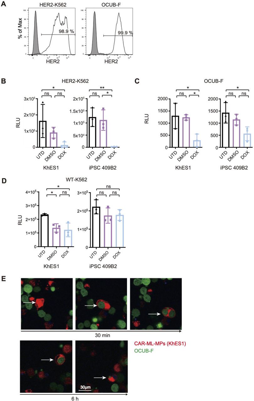 Human Her2 Erbb2 Protein, supplied by Sino Biological, used in various techniques. Bioz Stars score: 95/100, based on 1 PubMed citations. ZERO BIAS - scores, article reviews, protocol conditions and more https://www.bioz.com/product/biotinylated+human+erbb2/Human+HER2+%2F+ErbB2+Protein+(His+%26+AVI+Tag)%2C+Biotinylated/bio_rxiv__2024__07__22__604686-49-11-16 Average 95 stars, based on 1 article reviews
human her2 erbb2 protein - by Bioz Stars,
2026-09
95/100 stars
|
Buy from Supplier |
|
Sino Biological
recombinant human her2 protein  Recombinant Human Her2 Protein, supplied by Sino Biological, used in various techniques. Bioz Stars score: 95/100, based on 1 PubMed citations. ZERO BIAS - scores, article reviews, protocol conditions and more https://www.bioz.com/product/biotinylated+human+erbb2/Human+HER2+%2F+ErbB2+Protein+(Fc+Tag)%2C+Biotinylated/pmc10992513-117-7-12 Average 95 stars, based on 1 article reviews
recombinant human her2 protein - by Bioz Stars,
2026-09
95/100 stars
|
Buy from Supplier |
|
Sino Biological
human her2 / erbb2 protein (his & avi tag), biotinylated Human Her2 / Erbb2 Protein (His & Avi Tag), Biotinylated, supplied by Sino Biological, used in various techniques. Bioz Stars score: 95/100, based on 1 PubMed citations. ZERO BIAS - scores, article reviews, protocol conditions and more https://www.bioz.com/product/biotinylated+human+erbb2/Human+HER2+%2F+ErbB2+Protein+(His+%26+AVI+Tag)%2C+Biotinylated/custom%4010004-h27h-b%4010%2E21769%2FBioProtoc%2E5427 Average 95 stars, based on 1 article reviews
human her2 / erbb2 protein (his & avi tag), biotinylated - by Bioz Stars,
2026-09
95/100 stars
|
Buy from Supplier |
|
The Human ErbB2 Her2 Trastuzumab Biotinylated Antibody from R D Systems is a human monoclonal antibody to ErbB2 Her2 This antibody reacts with human The Human ErbB2 Her2 Trastuzumab Biotinylated Antibody has been validated for
|
Buy from Supplier |
|
Human HER2 /ErbB2 /CD340 Protein, Biotinylated (Biotinylated rhHER2) Thr 23 - Thr 652 (AAA75493), was labeled with biotin based on rh HER2 Protein, which was produced in human 293 cells (HEK293).Human Epidermal growth factor Receptor
|
Buy from Supplier |
Image Search Results
Journal: mAbs
Article Title: Beyond bispecificity: Controlled Fab arm exchange for the generation of antibodies with multiple specificities
doi: 10.1080/19420862.2021.2018960
Figure Lengend Snippet: Generation of multispecific DB-VHH constructs. Trastuzumab IgG1-K409R mAb (anti-HER2) C-termini are fused to VHHs directed against EGFR, IL6R or NKG2D via a GS-linker. CS06 IgG1-F405L mAb (anti-c-MET) is fused to the same VHH molecules in the same manner. After recombinant production and purification, parental IgG-VHHs are recombined pairwise by reduction and reoxidation. The matching K409R and F405L mutations drive the generation of heterodimeric multispecific DB-VHHs.
Article Snippet: The biosensors were then rinsed in kinetics buffer (KB; PBS + 0.1% Tween-20 + 1% bovine serum albumin (BSA)) for 45 s (baseline) and the corresponding antigen was associated to the biosensor for 300 s. Different antigen concentrations (1:2 dilution rows in KB) were tested in order to identify the dynamic range of the binding kinetics and the most suitable concentration ranges were determined as follows:
Techniques: Construct, Recombinant, Purification

Journal: mAbs
Article Title: Beyond bispecificity: Controlled Fab arm exchange for the generation of antibodies with multiple specificities
doi: 10.1080/19420862.2021.2018960
Figure Lengend Snippet: Binding affinities measured for each paratope using recombinant antigens
Article Snippet: The biosensors were then rinsed in kinetics buffer (KB; PBS + 0.1% Tween-20 + 1% bovine serum albumin (BSA)) for 45 s (baseline) and the corresponding antigen was associated to the biosensor for 300 s. Different antigen concentrations (1:2 dilution rows in KB) were tested in order to identify the dynamic range of the binding kinetics and the most suitable concentration ranges were determined as follows:
Techniques: Binding Assay, Recombinant, Construct

Journal: mAbs
Article Title: Beyond bispecificity: Controlled Fab arm exchange for the generation of antibodies with multiple specificities
doi: 10.1080/19420862.2021.2018960
Figure Lengend Snippet: Biolayer interferometry analysis of simultaneous antigen binding of tri- and tetraspecific DB-VHHs. (a), (b) and (c) show exemplary sensorgrams for trispecific molecules and (d) a tetraspecific DB-VHH. The first association step represents binding of the DB-VHH (200 nM) via its CS06 paratope to biotinylated c-MET immobilized to streptavidin biosensors. Second (and third for (D)) association step is performed using an IL6R, EGFR or NKG2D recombinant protein (200 nM). The last association step is performed using HER2 (200 nM). Kinetic buffer (KB) controls were applied as negative controls for each association step.
Article Snippet: The biosensors were then rinsed in kinetics buffer (KB; PBS + 0.1% Tween-20 + 1% bovine serum albumin (BSA)) for 45 s (baseline) and the corresponding antigen was associated to the biosensor for 300 s. Different antigen concentrations (1:2 dilution rows in KB) were tested in order to identify the dynamic range of the binding kinetics and the most suitable concentration ranges were determined as follows:
Techniques: Binding Assay, Recombinant

Journal: mAbs
Article Title: Beyond bispecificity: Controlled Fab arm exchange for the generation of antibodies with multiple specificities
doi: 10.1080/19420862.2021.2018960
Figure Lengend Snippet: Specific cell clustering due to simultaneous binding of the DB-VHHs to three different cancer cell lines. Flow cytometry cytograms represent the fluorescence signals of the different cell populations. (a) Cells without antibody construct. Upper left gate = HCC-1954 (HER2 +++ ) cells stained with DeepRed, lower left gate = MDA-MB-468 (EGFR +++ ) cells stained with CMRA, lower right gate = EBC-1 (c-MET ++ ) cells stained with CFSE, upper right gate = HCC-1954 + EBC-1 cell doublets. Events in all three fluorescence channels (cell triplets) were marked in red. Cells were incubated in the presence of 1 nM (b) bispecific DB, (c) and (d) trispecific DB-VHHs, (e) and (f) tetraspecific DB-VHHs.
Article Snippet: The biosensors were then rinsed in kinetics buffer (KB; PBS + 0.1% Tween-20 + 1% bovine serum albumin (BSA)) for 45 s (baseline) and the corresponding antigen was associated to the biosensor for 300 s. Different antigen concentrations (1:2 dilution rows in KB) were tested in order to identify the dynamic range of the binding kinetics and the most suitable concentration ranges were determined as follows:
Techniques: Binding Assay, Flow Cytometry, Fluorescence, Construct, Staining, Incubation

Journal: mAbs
Article Title: Beyond bispecificity: Controlled Fab arm exchange for the generation of antibodies with multiple specificities
doi: 10.1080/19420862.2021.2018960
Figure Lengend Snippet: Simultaneous interaction of the DB-VHHs with HCC-1954 and EBC-1 target cells and binding of recombinant IL6R. Flow cytometry cytograms represent the fluorescence signals of the two cell populations and bound recombinant IL6R, detected with an anti-His6 detection antibody. (a) Cells without antibody construct. Upper left gate = HCC-1954 (HER2+++) cells stained with DeepRed, lower right gate = EBC-1 (c-MET++) cells stained with CFSE, upper right gate = HCC-1954 + EBC-1 cell doublets. Events in all three fluorescence channels (HCC-1954 + EBC-1 + bound recombinant IL6R-His Tag) were marked in blue. Cells were incubated in the presence of 10 nM (b) bispecific DB, (c) and (d) trispecific DB-VHHs, (e) and (f) tetraspecific DB-VHHs.
Article Snippet: The biosensors were then rinsed in kinetics buffer (KB; PBS + 0.1% Tween-20 + 1% bovine serum albumin (BSA)) for 45 s (baseline) and the corresponding antigen was associated to the biosensor for 300 s. Different antigen concentrations (1:2 dilution rows in KB) were tested in order to identify the dynamic range of the binding kinetics and the most suitable concentration ranges were determined as follows:
Techniques: Binding Assay, Recombinant, Flow Cytometry, Fluorescence, Construct, Staining, Incubation

Journal: mAbs
Article Title: Beyond bispecificity: Controlled Fab arm exchange for the generation of antibodies with multiple specificities
doi: 10.1080/19420862.2021.2018960
Figure Lengend Snippet: DB-VHHs elicit potent and specific NK cell-mediated target cell killing. Tumor cells were incubated with primary effector cells (NK cells) at a 1:5 ratio in the presence of antibody constructs in different concentrations. Error bars represent standard deviation of two biological replicates. Wildtype CS06 and trastuzumab with active Fc effector functioning were used as an ADCC reference (green). (a) and (b): c-MET-positive EBC-1 target cells, (c) and (d) HER2-overexpressing SK-BR-3 target cells. (a) and (c): NK cell cytotoxicity triggered by parental antibodies. (b) and (d): DB-VHHs serve as NK cell engager and mediate tumor cell killing.
Article Snippet: The biosensors were then rinsed in kinetics buffer (KB; PBS + 0.1% Tween-20 + 1% bovine serum albumin (BSA)) for 45 s (baseline) and the corresponding antigen was associated to the biosensor for 300 s. Different antigen concentrations (1:2 dilution rows in KB) were tested in order to identify the dynamic range of the binding kinetics and the most suitable concentration ranges were determined as follows:
Techniques: Incubation, Construct, Standard Deviation

Journal: mAbs
Article Title: Beyond bispecificity: Controlled Fab arm exchange for the generation of antibodies with multiple specificities
doi: 10.1080/19420862.2021.2018960
Figure Lengend Snippet:
Article Snippet: The biosensors were then rinsed in kinetics buffer (KB; PBS + 0.1% Tween-20 + 1% bovine serum albumin (BSA)) for 45 s (baseline) and the corresponding antigen was associated to the biosensor for 300 s. Different antigen concentrations (1:2 dilution rows in KB) were tested in order to identify the dynamic range of the binding kinetics and the most suitable concentration ranges were determined as follows:
Techniques: Variant Assay, Hydrophobic Interaction Chromatography, Mutagenesis, Molecular Weight, High Performance Liquid Chromatography
Journal: bioRxiv
Article Title: Enhanced anti-tumor effects through continuous administration of engineered CAR-macrophages derived from pluripotent stem cell-derived myeloid cell lines
doi: 10.1101/2024.07.22.604686
Figure Lengend Snippet: (A) Representative histograms showing HER2 expression in HER2-K562 and OCUB-F. (B, C) Cytotoxicity assay of CAR-ML-MPs derived from KhES1 and iPSC 409B2 strains against HER2-K562 (B) and OCUB-F (C). Effector-to-target ratio were 5:1 to HER2-K562 and 3:1 to OCUB-F, respectively (n = 3, independent experiment). UTD indicates untreated group. DMSO and DOX represent conditions treated without and with induction of CAR expression, respectively. RLU: relative light unit. Dunnett’s tests were performed as an a priori multiple comparison. *P < 0.05, **P < 0.01. Error bars represent mean ± standard deviation (SD). (D) Cytotoxicity assay of CAR-ML-MPs against wild type K562. (E) Results of time-lapse imaging of co-culture of CAR-ML-MPs (KhES1 strain) and OCUB-F (E/T ratio 1:1) after 30 minutes and 6 hours. Representative ML-MP cell (white arrow) showed phagocytosis of OCUB-F. See Video S1.
Article Snippet: For confirmation of anti-HER2 CAR expression, primary staining was performed with
Techniques: Expressing, Cytotoxicity Assay, Derivative Assay, Comparison, Standard Deviation, Imaging, Co-Culture Assay

Journal: bioRxiv
Article Title: Enhanced anti-tumor effects through continuous administration of engineered CAR-macrophages derived from pluripotent stem cell-derived myeloid cell lines
doi: 10.1101/2024.07.22.604686
Figure Lengend Snippet: (A) Experimental time course of in vivo assays comparing single and consecutive tumor treatments. (B) Bioluminescence images and (C) quantitative values of bioluminescence from Luc-HER2-K562 cells at the indicated days and conditions. Statistical analysis was performed using a priori Dunnett’s multiple comparisons test. UTD indicates untreated group; 1 and 1+6 indicate single treatment alone and with additional treatment, respectively. *P < 0.05, **P < 0.01. Error bars represent mean ± standard deviation (SD). (D) Kaplan–Meier plot showing the survival of each group (n = 3 for each group). Statistical analysis was performed with Log-rank (Mantel–Cox) test. (E) Experimental time course of repeated Luc-ML-MP transplantation assay. Bioluminescence images of (F) Luc-ML-MPs and (G) their quantification (n = 3). Error bars represent mean ± standard deviation (SD).
Article Snippet: For confirmation of anti-HER2 CAR expression, primary staining was performed with
Techniques: In Vivo, Standard Deviation, Transplantation Assay

Journal: bioRxiv
Article Title: Enhanced anti-tumor effects through continuous administration of engineered CAR-macrophages derived from pluripotent stem cell-derived myeloid cell lines
doi: 10.1101/2024.07.22.604686
Figure Lengend Snippet: (A) Schematic diagram of DOX-inducible anti-HER2 CAR construct. The construct includes HER2 recognition domain, intracellular hinge, CD28 and CD3ζ. TRE: tetracycline-response element. EF1: EF1 Alpha Promoter. TetR: Tet Repressor. ITR: inverted terminal repeat. (B) Proliferation curve of PSC-MLs derived from CAR-PSCs. On the left is mean ± standard deviation (SD) data for one passaging culture (n = 3, independent experiment). On the right, growth curves of KhES1-derived MLs cultured for 60 days. (C) Morphology of ML-MPs. Phase-contrast images (Upper panels) and May–Giemsa staining images (Lower panels). (D, E) Representative histograms showing macrophage cell-surface markers on CAR-ML-MPs. Panels (D) and (E) depict data from iPSC 409B2 and KhES1 strains, respectively. The rightmost panel displays histograms showing anti-HER2 CAR expression on the surface of CAR-ML-MPs in the presence of DOX. Gray fills represent isotype controls in all panels. (F) Expression of OX40L (left two panels) and 4-1BBL (right two panels). The relative quantity (Rq) of RNA expressions was measured in ML-MPs derived from the iPSC 409B2 strain (left) and the KhES1 strain (right) under both conditions, with and without DOX, compared to undifferentiated cells.
Article Snippet: For confirmation of anti-HER2 CAR expression, primary staining was performed with
Techniques: Construct, Derivative Assay, Standard Deviation, Passaging, Cell Culture, Staining, Expressing

Journal: bioRxiv
Article Title: Enhanced anti-tumor effects through continuous administration of engineered CAR-macrophages derived from pluripotent stem cell-derived myeloid cell lines
doi: 10.1101/2024.07.22.604686
Figure Lengend Snippet: (A) Experimental time course of in vivo tumor treatment assay. (B, C) Bioluminescence images (B) and quantitative values of bioluminescence (C) from Luc-HER2-K562 cells at the indicated days and conditions. Statistical analysis was performed using a priori Dunnett’s multiple comparisons test. UTD indicates untreated group. DMSO and DOX represent conditions treated without and with induction of CAR expression, respectively. *P < 0.05, ***P < 0.001. Error bars represent mean ± standard deviation (SD). (D) Kaplan–Meier plot showing the survival of each group (n = 5 for each group). Statistical analysis was performed with Log-rank (Mantel-Cox) test. (E) Experimental time course of Luc-ML-MP transplantation assay. Bioluminescence images of (F) Luc-ML-MPs and (G) their quantification (n = 3). Error bars represent mean ± standard deviation (SD).
Article Snippet: For confirmation of anti-HER2 CAR expression, primary staining was performed with
Techniques: In Vivo, Expressing, Standard Deviation, Transplantation Assay
Journal: Cancer Immunology, Immunotherapy : CII
Article Title: Chimeric antigen receptor clustering via cysteines enhances T-cell efficacy against tumor
doi: 10.1007/s00262-022-03195-4
Figure Lengend Snippet: Cysteines in the hinge facilitate antigen-triggered CAR cluster formation and promote CAR T-cell activity A Gene schematics of CARCys and CARSer targeting HER2. B Human T cells derived from healthy donors were transduced with or without HER2-CARCys or HER2-CARSer mRNA to generate NT or HER2-CAR-T cells. Twelve hours post-transduction, the CAR expression on these T cells was detected by flow cytometry. Data were representative of at least three independent experiments. C–E CARCys and CARSer distribution on these T cells was observed using STORM system after stimulation with HER2 antigen for 30 min. The tables containing x × y co-ordinates of each CAR molecule were obtained from STORM, and DBSCAN implemented in MATLAB was used for CAR clusters identification. For each T cell, a 5 × 5 μm2 ROI in cell center was selected for further analysis of cluster number and diameter by DBSCAN. C The representative cluster maps of CARCys and CARser on primary human T cells. D The number of CAR clusters per ROI. E The diameters of CAR clusters. Data shown as mean ± SEM were from 13 to 16 cells of healthy donors. F CARCys and CARSer-T cells were co-cultured with HER2+N87 or HER2−K562 tumor cells. Four hours later, CD107a translocation of CAR-T cells was detected by flow cytometry. Data shown as mean ± SEM were pooled from three healthy donors. G, H CARCys and CARSer-T cells were co-cultured with HER2+N87 at indicated E: T ratios for 6 or 48 h. The cytotoxicity of CAR-T cells was calculated by the target cells’ viability using CCK8. Data shown as mean ± SEM were from one of two independent experiments using two healthy donors. P values were determined by unpaired Student’s t test in (D, E), and two-way ANOVA in F, G, and H. **P < 0.01, ***P < 0.001
Article Snippet: CAR-T cells were stimulated with 2 μg/mL
Techniques: Activity Assay, Derivative Assay, Transduction, Expressing, Flow Cytometry, Cell Culture, Translocation Assay

Journal: Cancer Immunology, Immunotherapy : CII
Article Title: Chimeric antigen receptor clustering via cysteines enhances T-cell efficacy against tumor
doi: 10.1007/s00262-022-03195-4
Figure Lengend Snippet: CARCys-T cells exhibit increased antigen-specific activity compared to CARconv-T cells in vitro Human T cells derived from healthy donors were transduced with or without HER2-CARconv or HER2-CARCys mRNA to generate NT or HER2-CAR-T cells. A These CAR-T cells were stimulated with HER2 antigen for 30 min, and identified the antigen-triggered CAR clusters by STROM and DBSCAN analysis. B, C The number of CAR clusters per ROI (5 × 5 mm2) and the diameter of CAR clusters were calculated by DBSCAN analysis. Data shown as mean ± SEM were from 10 CAR-T cells of healthy donors. D Twelve hours post-transduction, NT- or CAR-T cells were cultured in Opti-MEM for 6 h to reduce background. These T cells were incubated with biotinylated HER2 protein (2.5 μg/mL) for 30 min at 4 °C and washed twice with cold PBS. Then, these T cells were cross-linked with streptavidin (5 μg/mL) at 37 °C for 30 s to measure the phosphorylation of CD3ζ and ZAP70, and cross-linked for 2 min to detect the phosphorylation of ERK1/2, S6 and AKT. E The enriched gene sets of CARconv-T cell comparing with CARCys-T cells after 10-h antigen stimulation. F The heat map of selected differentially expressed genes related to T cell activation, cytokine production, cytotoxicity, and chemokine signaling. Differentially expressed genes were selected with an adjusted P value (false discovery rate, FDR) < 0.05 and absolute fold change ≥ 2. G–J NT, CARconv, or CARCys-T cells were co-cultured with HER2−K562 or HER2+N87. (G) CD25 and Ki67 expressions of T cells were detected by flow cytometry 24 and 48 h post-co-culture, respectively. Data shown as mean ± SEM were pooled from four healthy donors. H, I IFN-γ and IL-2 production of T cells were measured by ELISA 24 h post-co-culture. Data shown as mean ± SEM were from one of three independent experiments using three healthy donors. J Cytotoxic activity of T cells was measured by performing CCK8 assay 12 h post-co-culture at indicated E: T ratios; data shown as mean ± SEM were from one of two independent experiments using two healthy donors. K–M Human T cells were transduced with HER2-CARconv, HER2-CARCys or HER2-CARSer mRNA to generate CAR-T cells. Twelve hours post-transduction, these CAR-T cells were treated with 10 mM/mL DTT and co-cultured with HER2+N87 tumor cells at the ratio of 1: 2. Six hours later, the CD107a translocation (K), the TNF-α (L) and IFN-γ (M) production were detected by flow cytometry. Data shown as mean ± SEM were from two independent experiments. P values were determined by unpaired Student’s t-test in (C), one-way ANOVA in (G), and two-way ANOVA in (H–M). *P < 0.05, **P < 0.01, and ***P < 0.001
Article Snippet: CAR-T cells were stimulated with 2 μg/mL
Techniques: Activity Assay, In Vitro, Derivative Assay, Transduction, Cell Culture, Incubation, Activation Assay, Flow Cytometry, Co-Culture Assay, Enzyme-linked Immunosorbent Assay, CCK-8 Assay, Translocation Assay

Journal: Cancer Immunology, Immunotherapy : CII
Article Title: Chimeric antigen receptor clustering via cysteines enhances T-cell efficacy against tumor
doi: 10.1007/s00262-022-03195-4
Figure Lengend Snippet: CARCys-T cells with different CAR framework exhibit stronger antigen-specific activity than CARconv-T cells. Human T cells derived from healthy donors were transduced with or without second-generation HER2-CARconv or HER2-CARCys with a 4-1BB costimulatory domain, or third-generation CD19-CARconv or CD19-CARCys mRNA to generate NT- or CAR-T cells. A Twelve hours post-transduction, HER2-CAR expression levels of NT-, HER2-CARconv-, or HER2-CARCys-T cells were detected by flow cytometry. B NT-, HER2-CARconv- or HER2-CARCys-T cells were co-cultured with HER2−K562 or HER2+N87. Twenty-four hours post-co-culture, IFN-γ production of these T cells were measured by ELISA. Data shown as mean ± SEM were from one of two independent experiments using two healthy donors. C CD107a translocation of NT-, HER2-CARconv-, or HER2-CARCys-T cells was detected by flow cytometry four hours post-co-culture. D Twelve hours post-co-culture, the cytotoxicity of NT-, HER2-CARconv-, or HER2-CARCys-T cells was analyzed by performing CCK8 assay after at indicated E: T ratios. Data shown as mean ± SEM were from one of two independent experiments using two healthy donors. E Twelve hours post-transduction, CD19-CAR expression levels of NT-, CD19-CARconv-, or CD19-CARCys-T cells were detected by flow cytometry. F, G NT-, CD19-CARconv-, or CD19-CARCys-T cells were co-cultured with CD19+Ramos for 12 h. CD25, CD69, CD95, and Perforin expression levels and cytokine production (TNF-α, IFN-γ) of these T cells were detected by flow cytometry. H NT-, CD19-CARconv-, or CD19-CARCys-T cells were co-cultured with CD19− K562 or CD19+ Ramos. CD107a translocation of NT, CD19-CARconv, or CD19-CARCys-T cells was detected by flow cytometry four hours post-co-culture. Data shown as mean ± SEM were pooled from two healthy donors. P values were determined by two-way ANOVA in (B, D, H). *P < 0.05, and **P < 0.01
Article Snippet: CAR-T cells were stimulated with 2 μg/mL
Techniques: Activity Assay, Derivative Assay, Transduction, Expressing, Flow Cytometry, Cell Culture, Co-Culture Assay, Enzyme-linked Immunosorbent Assay, Translocation Assay, CCK-8 Assay